Structure of the excavata, chromalveolata, and Amoebozoa
These members of the microbial eukaryotes are unicellular, heterotrophic, parasitic, symbiotic or photosynthetic organisms. They may be motile or nonmotile. They contain many of the organelles found in other eukaryotes, including nuclei, mitochondria (absent in some groups), chloroplasts (absent in some groups), ribosomes, endoplasmic reticulum (ER), Golgi vesicles, microtubules, and microfibrils.
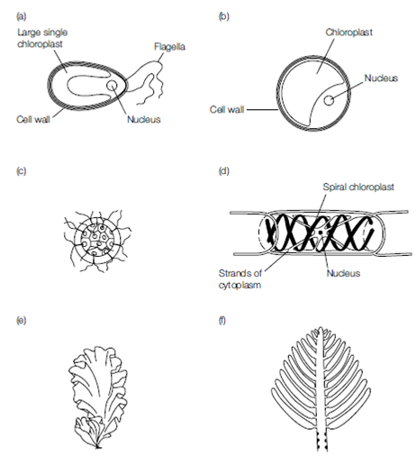
Figure Chlorophytan cells and thalli. (a) Motile unicell; (b) nonmotile unicell; (c) colonial forms; (d) filamentous forms; (e) membranous (only two cells thick); and (f) tubular. Courtesy of CJ Clegg.
Chloroplasts can be very variable in this group, and their shape and pigment content are useful distinguishing taxonomic features. The numbers of ER membranes that surround the chloroplast, and the number of thylakoid stacks within them, are also important indicators of its symbiotic origin. Unique organelles found in the protista include contractile vacuoles that control the influx of water to the cells due to osmosis. Some of the protista have hydrogenosomes where fermentative reactions take place. Some excavates and chromalveolates have a cell covering of scales or plates formed of pseudochitin, and this may be impregnated with calcium or silica scales. These scales are secreted from the Golgi bodies within alveoli. Some species have the ability to form thick-walled cysts during a resting phase in the life cycle. These cysts can be very resistant to adverse conditions.
This group of organisms is characterized by a considerable variation in morphology. Free-floating species are radially or laterally symmetrical and streamlined into bullet or kidney shapes. At its simplest, the outer surface of the cells may be simply the plasma membrane. This is seen in the gametes of some species, in amebae, and in some of the intracellular parasites. Such a ‘naked’ cell has advantages in nutrient uptake that
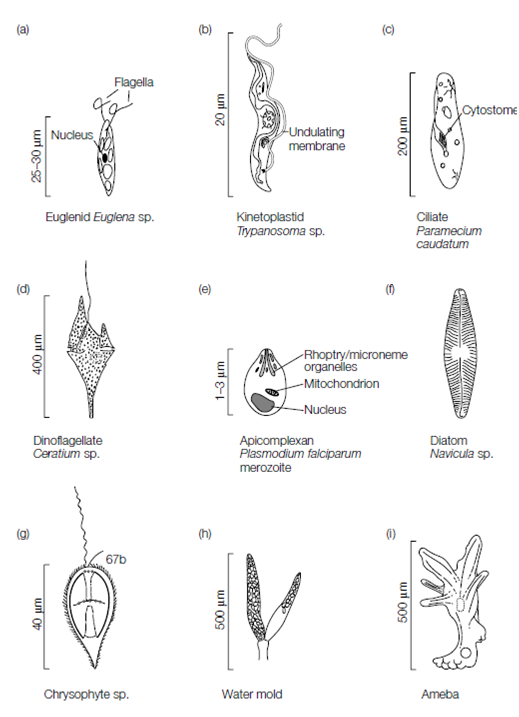
Figure Structure of the Excavata and Chromalveolata.
outweigh its vulnerability. The naked membrane can be extended with lobes of cytoplasm called pseudopodia that are important in locomotion and feeding. This type of movement requires that the cell is in contact with a solid surface.
The cell is partitioned into two regions, the viscous ectoplasm and the more liquid endoplasm. Within the ectoplasm are myosin and actin filaments, while in the endoplasm only unpolymerized actin is present. To produce ameboid movement the microfilaments in the ectoplasm produce pressure on the endoplasm moving the endoplasm to one part of the cell, and away from another.
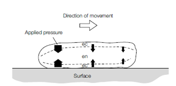
Figure A proposed mechanism for ameboid movement. ec, ectoplasm; en, endoplasm.
Motility can also be via flagella or cilia that propel the cells through water by rhythmic beating. Flagella typically exhibit a sinusoidal motion in propelling water parallel to their axis. The undulating action of the flagellum either propels water away from the surface of the cell body or draws water towards and over the cell body. Cilia exhibit an oar-like motion, propelling water parallel to the cell surface.