Reciprocal inhibition
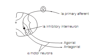
Axon collaterals of Ia afferents from muscle spindles synapse with glycinergic Ia inhibitory interneurons in lamina VII (IaINs) which project to motor neurons of antagonist muscles. This disynaptic circuit permits antagonist muscles to be relaxed during agonist contraction as shown in figure. The inhibition of equally antagonistic muscle motor neurons is known as reciprocal inhibition.
IaIN activity mediating reciprocal inhibition is customized by descending motor pathways (rubrospinal, corticospinal, and vestibulospinal) and locomotor networks in the cord.
This occurs:
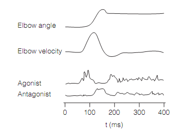
- To facilitate quick movements. Since muscle contractions are thus long lasting, muscles follow loyally only slowly changing neural input. Quick changes in motor neuron firing cannot be translated into equivalent modifications in muscle tension. Therefore to produce rapid fluctuations in tension the motor system modifies contraction in agonist and antagonist muscles. This is helped by the reciprocal inhibition as shown in figure.

Figure: A disynaptic reflex for reciprocal inhibition of antagonist muscle.
- To adjust muscle stiffness therefore it is suitable for the load. The rigidity of a joint is increased by co-contraction of muscles with opposing actions at the joint. For illustration, co-contraction of the biceps and triceps increase stiffness of the elbow. Given one muscle contracts more strongly than the other i.e., the joint moves. Co-contraction stabilizes a joint, it gives better control whenever loads change suddenly as a given difference among expected and actual load will have a smaller effect on limb trajectory if a joint is stiffer. Co-contraction needs suppression of reciprocal inhibition.

Figure: Pattern of activation of agonist (biceps) and antagonist (triceps) muscles to facilitate quick movement (i.e., elbow flexion).