Sleep pathways
Monoaminergic neurons in the ascending reticular formation needs to generate the awake state and to increase arousal and the responsiveness of the cortex to sensory input constitute an ascending arousal system. It splits into two branches at the stages of the diencephalon. One branch projects by the lateral hypothalamus to the cerebral cortex from a variety of sources that are:
- Noradrenergic neurons of locus coeruleus
- Serotonergic cells in raphe nuclei
- Histaminergic neurons in TMN (abbreviate as tuberomammillary nucleus) of the hypothalamus
This branch is augmented through cholinergic neurons in the basal forebrain.The serotonergic and noradrenergic neurons fire at the highest rate in alert animals, have low firing rates during NREM sleep and go silent during REM sleep therefore are called wake-on or REM-off cells.
The other second branch projects to the thalamus and contains are as follows:
- Histaminergic neurons in the TMN that make excitatory synapses with thalamic relay neurons
- Cholinergic neurons in the pons which is PPN, lateral dorsal tegmental nucleus, and pedunculopontine nucleus LdT that inhibit GABAergic thalamic reticular neurons and therefore excite thalamic relay cells
The pontine cholinergic cells are active in the during of REM sleep and wakefulness and are responsible for the desynchronization of the EEG in these states—but become quiescent during NREM sleep and therefore are described as wake-on or REM-on cells.Neurons that use orexins, located in the tuberal region of the hypothalamus excite all the groups of neurons listed above and are active during the REM sleep and awake state. Orexigenic neurons probably stabilize whichever state (awake or REM) the brain is in.
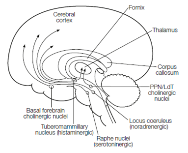
Figure: Ascending arousal system.
Only 5–10 percent of synapses in the thalamus are made through afferent terminals. Many of the thalamus is concerned with controlling that sensory information is sent to the cortex (attention). Thalamic relay cells project to get reciprocal connections and the cortex from the cortex in Figure. Relay cells and cortical projection both neurons use glutamate and are excitatory. Specific sensory input from the retina directly excites the relay cells. The relay cells are topic to inhibition from GABAergic interneurons and from the thalamic reticular nucleus (TRN) a sheet of GABAergic neurons covering the thalamus.
There are two modes of thalamic relay neurons of firing that are:
- The relay cells are depolarized through input from the ascending arousal system while tonic firing of single action potentials occurs in the awake state. In this mode, transmission of sensory input from thalamus to cortex takes place.