Horizontal cells and lateral inhibition
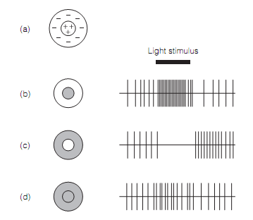
Lateral inhibition improves contrast and is brought about by horizontal cells. The lateral inhibition can be seen in receptive fields (RFs) of both bipolar and ganglion cells. These are circular and separated into an inner center and an outer surround. The stimulation of these two areas individually produces opposite effects on the cells. In the case of ganglion cell, for illustration as shown in figure, background firing rate is increased by center illumination but silenced by light on the surround. Whenever light fills the entire receptive field there is little change in the firing rate. Off ganglion cell RFs have the converse response, with center illumination generating inhibition and surround illumination excitation.

Figure: Extracellular recording from on ganglion cells: (a) receptive field; (b) central illumination; (c) surround illumination; (d) overall illumination.
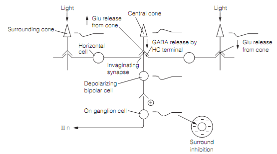
Lateral inhibition occurs as horizontal cells form reciprocal connections at triad synapses among neighboring photoreceptors. The details are a bit complex. In the dark, horizontal cells are excited by glutamate discharge from photoreceptors; however they discharge GABA that tends to inhibit the photoreceptors. The light that hyperpolarizes surrounding photoreceptors causes them to secrete less glutamate therefore reducing horizontal cell excitation. This means that GABA discharge from the horizontal cells is lowered, which in return permits the central cone to depolarize somewhat, hence it releases more glutamate. The last step in the series depends exactly on the kind of bipolar cell the central cone synapses with. When it is a depolarizing (on) bipolar cell the increased glutamate will cause it to hyperpolarize, since glutamate is inhibitory at invaginating synapses. When it is a hyperpolarizing (off) bipolar cell the increased glutamate will make the bipolar cell depolarize. Note that in each situation the bipolar cell response (and so that of the ganglion cell with which it synapses) is the opposite for surround compared with central illumination as shown in figure below:

Figure: The mechanism of lateral inhibition by horizontal cells
The horizontal cells are extensively interconnected through gap junctions forming a network that spans a region of retina known as the S space. The S space horizontal cells give the signal for surround inhibition and it is thought that this signal is a measure of the mean luminance over quite a broad region of retina.