Ganglion cells
The axons of ganglion cells form the optic nerve. Only ganglion cells are the retinal cells able of firing the action potentials. There are three ganglion cell kinds. The Parvocellular (P) ganglion cells are small and by far the most numerous with about a million in each retina. The Magnocellular (M) ganglion cells are large and number about 100 000 per retina. The Koniocellular (K) ganglion cells are small and look like P cells physiologically.
The P and M types differ in many significant respects as follows:
- The P cells have smaller RFs as compare to M cells.
- Since of their smaller size, the P cells have a slower conduction velocity as compare to M cells.
- Whereas P cells frequently show sustained responses, the M cells respond transiently to a pro-longed visual stimulus.
- The P cells are generally wavelength selective while M cells are not.
- The M cells are very much sensitive as compare to P cells to low-contrast stimuli.
From such differences it can be inferred that P cells should get their input from single cones or from numerous cones with similar wavelength sensitivity (S, M, or L). By contrast, the M cells get input from M and L cones altogether (however not S cones), and from rods. Therefore P, but not M, cells should be included in the color vision. The fast, transient responses of M cells make them adapted for the motion detection. The small RFs of P cells and their continuous responses are appropriate for fine form discrimination. The different functional properties of P, M, and K ganglion cells is the starting point for parallel processing in the visual system.
The P cells are color single opponent cells. They have RFs which are excited by one kind of cone cell but inhibited by the other. The two kinds of P cell can be distinguished by the nature of their RFs as shown in figure. The most general are the red–green cells in which the RF compares input from M and L cones. The Blue–yellow cells do not have a center–surround pattern but are either excited by S cones or reserved by a combined M plus L cone signal, or vice-versa. They are known as blue–yellow cells since combining the inputs of M and L cones provides the sensation of yellow.
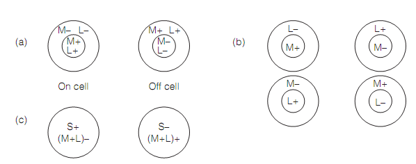
Figure: Receptive fields of retinal ganglion cells: (a) M ganglion cells; (b) concentric single opponent (red–green) P cells; (c) coextensive single opponent (blue–yellow) P cells.
The red–green cells respond in a different way to small or large spots of light. For illustration, a green-on/red-off cell will be uniformly stimulated by a small green or white spot which covers the center of the RF. This is as white light consists of the wavelengths that excite the green cones. Though, similar cell will be excited by a large green spot but not by a large white spot. This is as the white light consists of the wavelengths that stimulate the red-off surround. A large red spot will silence the cell. Therefore, in general, such cells are more wavelengths selective for large stimuli than the small ones. For small spots, they cannot differentiate red or green from white light, but they can signal the brightness.
The M cells get combined input from two kinds of cones therefore they are known as broad band cells and their RFs measure only brightness contrast; that is the M cells are color blind.A few K cells are blue-on cells; others are motion sensitive and have low spatial resolution.