Gel electrophoresis:
When a DNA molecule is cut through a restriction enzyme the DNA fragments that's called restriction fragments from which restriction digest can be divided through gel electrophoresis shown in the figure. Electrophoresis on a polyacrylamide gel will divide small DNA fragments of less than about 500 bp in size, but agarose gels (that have larger pores) are required to separate huger DNA fragments. The DNA digest divided into a series of bands representing the restriction fragments. Still little fragments travel further in the gel than larger fragments, the size of every fragment can be determined through measuring its migration distance relative to standard DNA fragments of known size. The DNA can be located over gel electrophoresis through staining with ethidium bromide which binds to the DNA and fluoresces a bright orange. In the other words, if the DNA is labeled with a radioisotope like as 32P, the bands can be detected after electrophoresis through laying the gel against an X-ray film whereby the radioactivity causes silver grains to be formed in the film emulsion providing black images corresponding to the radioactive bands (autoradiography).
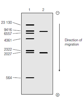
Figure: Agarose gel electrophoresis of DNA fragments. DNA fragments of known size were electrophoresed in lane 1 (the sizes in bp are given on the left). A restriction digest of the sample DNA was electrophoresed in lane 2. By comparison with the migration positions of fragments in lane 1, it can be seen that the two sample DNA fragments have sizes of approximately 9000 bp and 2000 bp. The sizes could be determined more accurately by plot- ting the data from lane 1 as a standard curve of log DNA size vs. migration distance and then using this to estimate the size of the sample DNA fragments from their measured migration distances.