Cone cells
The Cone photoreceptors are very dense at the fovea and their numbers fall off sharply beyond 50 of it. They have a low sensitivity to light and do not saturate except in most intense light, therefore are used for the photopic (i.e., daylight) vision. The Photopic vision has high acuity as there is little or no convergence between cone cells and bipolar cells. The Cone cells integrate photon responses over a short time and therefore are able to solve a flicker frequency of less than of about 55 Hz.
At low light levels (example, dusk) rod cells operate beside the cones, boosting the cone cell signal by means of electrical synapses to preserve the color perception. This is known as mesopic vision.
There are three populations of cone cells that differ in their spectral sensitivity as shown in figure.
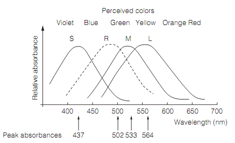
Figure: Spectral sensitivities of photoreceptors: Here, S=short-wavelength cones; R=rods; M=medium-wavelength cones; and L=long-wavelength cones.
However more appropriately called short (S), medium (M), and long (L) wavelength cones they are frequently referred to as blue, green, and red cones respectively, however their peak sensitivities are not best explained by such colors. The S cones are sensitive to wavelengths down to 315 nm. Though, the normal eye does not see wavelengths shorter than 400 nm (i.e., ultraviolet) as they are absorbed by the lens.
The color vision needs comparisons of the relative strengths in the outputs of the S, M, and L cones. The S cones constitute only about 5–10% of the total number of cones and are missing from the center of the fovea. This is as the lens suffers from chromatic aberration, in which the short-wavelength light is not brought to focus at similar point as longer wavelengths, causing image blurring. This would compromise the high acuity vision. Accordingly, color vision at the central fovea is dichromatic and moreover, M and L cones are distributed arbitrarily leaving patches in which there is only one population of cone. These features mean that the color vision is coarse grained and cannot resolve the fine detail.
The Scotopic vision is achromatic as all rod cells have similar spectral sensitivity curve. They are not capable to distinguish between wavelengths on the rising and falling limbs of the spectrum which excite the cell to the similar extent. Under scotopic vision the sensitivity of the eye is determined by the rod cells and peaks at around 500 nm. Under photopic situation the wavelength sensitivity is governed by cones and is maximal at 550 nm. This shift in wavelength sensitivity during the mesopic vision is known as the Purkinje shift. It means that as the dusk falls red fades then first and the last color to be lost is green.
Whenever moving from bright to very dim light the sensitivity of the retina to light increases a million-fold over a time period of 30 min or longer. This is known as the dark adaptation and is a property of the photoreceptors. The dark adaptation has two phases as shown in figure. The first is due to the cone cells that increase the sensitivity about 100 fold; the second longer phase is due to the rod cells.
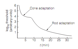
Figure: The time course of dark adaptation.
The light adaptation takes place whenever going from dim to brightly lit conditions. It is very much faster than the dark adaptation.