Extrastriate visual cortex
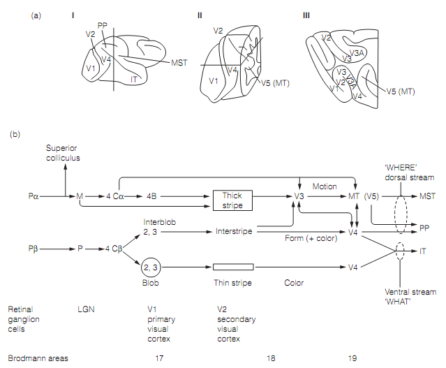
The segregation of visual information for motion, form, and color in V1 is preserved in the extrastriate visual cortex that is a word applied to the entire visual cortex except V1. The extrastriate cortex of primates holds about 30 regions that can be distinguished on the grounds of connections, cyto-architecture, and physiological properties. Most have a retinotopic map of a few aspect of the visual world. It involves not only occipital cortex regions 18 and 19 but also regions of parietal and temporal cortex. The position and connections of the major visual cortical regions are depicted in figure a and b respectively.
Most of the outcomes from V1 to V2, secondary visual cortex that occupies part of region 18. V2 explains a characteristic cytochrome oxidase staining pattern, irregular thick and thin stripes running at right angles to the V1/V2 border. Pathway tracing methods and electrophysiological studies reveal how the magnocellular and parvocellular pathways continue into V2 and beyond.
Cells in the V2 thick stripes are motion sensitive and binocular, being driven by a privileged retinal disparity. The thick stripe obtains inputs from layer 4 of the interblob area of V1 and sends much of its output, through V3, to the medial temporal (MT) visual cortex, V5. Human V5 lesions outcomes in loss of the capability to perceive motion. Therefore, the V2 thick stripe–V3–V5 (MT) connection is the extension of the magnocellular pathway as shown in figure (b), and concerned with the motion and depth perception.
The interstripe area of V2 gets its inputs from the V1 interblob areas (layers 2 and 3) and sends outputs to V3 and then to V4. Most cells in V3 and V4 are orientation selective.

Figure: Parallel processing in the visual system. (a) Anatomy of the visual regions in the macaque: (i) left cerebral hemisphere; (ii) coronal section via the posterior third of the hemisphere; (iii) horizontal section.
Hence this route is a continuation of the PI parvocellular pathway concerned primarily with form perception.
The blobs of V1 project to the thin stripe of V2 which in return sends outputs to visual regionV4. That this route is the extension of the PB parvocellular pathway for color vision is supported by the loss of color vision which takes place in patients with damage to human V4.
The M, PI, and PB pathways are not entirely independent. The reciprocal pathways exist among V3 and V4, and V5 and V4 which presumably permit interactions among M and PI systems, both of which add to stereopsis. The interaction of motion and form analysis is probably needed for the specification of moving objects. There seems to be no cross talk, though, among M and PB pathways. M system is color blind and for equiluminant stimuli (those changeable in color but not in brightness), that can only be perceived by the PB system, the perception of motion vanishes. Furthermore, though the PI system uses color contrast to localize borders, form information is not available to the PB pathway: whenever looking at equiluminant blocks of color they emerge to “jump around” as the PB system cannot localize boundaries.
During saccades the M, but not the P system, is shut down. Therefore the M (motion) system is not confused by the fast eye movements. The response times of the P system are adequately slow that they are not affected by the shifting image.