Spinal cord
The human spinal cord has around 108 neurons. A transverse section through the spinal cord displays a butterfly-shaped central gray matter that contains neuron cell bodies. The white matter surrounding the gray is mostly axons in ascending and descending tracts and gets its color from the high content of myelin. In the middle is the central canal that holds the cerebrospinal fluid (CSF), though in adults it is generally closed.
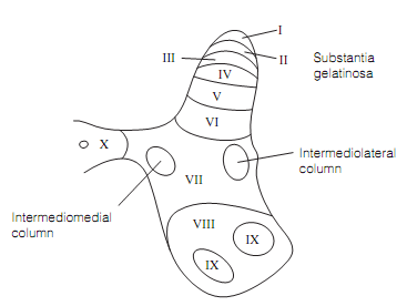
The Sensory axons enter spinal cord via the dorsal roots to synapse largely with cells in the dorsal horns of the spinal gray matter. Bigger diameter fibers enter more medially and extend more deeply into the dorsal horn than the smaller ones. The Motor neuron cell bodies lie in the ventral horns of the spinal gray and their axons exit through the ventral roots. In the spinal gray matter, 10 columns running throughout the cord can be distinguished on the basis of cell size. On transverse section these columns looks like as Rexed laminae as shown in figure.

Each and every lamina has distinctive input–output relations that reflect the functional specialization. And hence, while nociceptor afferents synapse on cells in lamina II, cutaneous mechanoreceptor afferents finish in deeper layers of the dorsal horn, and lamina VII and lamina IX house preganglionic autonomic neurons and motor neurons correspondingly.
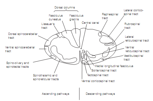
The white matter is organized into tracts identified by origin and destination. For illustration, the descending tract from the cerebral cortex is known as the corticospinal tract while the ascending path that terminates in the thalamus is the spinothalamic tract as shown in figure.

Figure: Pathways in the spinal cord white matter.