Brain
There are three most primary structural components to the brain:
- The tracts or pathways enter neuraxis at different levels, rise or low, and these, altogether with internal tracts that go from one part of the brain to the other, constitute the white matter.
- Nuclei embedded in the white matter are the clusters of neuron cell bodies. Several neural structures are composed of groups of nuclei. The thalamus, for illustration, consists of a few 30 nuclei.
- The two brain structures, the cerebrum and the cerebellum, are enclosed by cortex, a thin rind with a very high density of neuron cell bodies. In wiring words the cortex looks to be a simple circuit between a few neuron types, which repeat millions of times.
Altogether, the cortex and nuclei are the gray matter of the brain. The neural systems are comprised of interconnected nuclei and cortical regions which serve a common function. The visual system, for illustration, consists of the retinas, the lateral geniculate nuclei of the thalamus, the visual cortex and the pathways among them.The most basic division of the brain into midbrain, hindbrain, and forebrain can be discerned in the human embryo by the end of the 4th week whenever the CNS is a hollow neural tube which is as shown in figure.
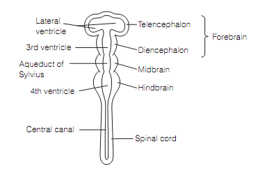
Figure: The human embryo neural tube, at 28 days gestation.
The hindbrain contains pons, medulla, and cerebellum; however the hindbrain (minus the cerebellum) and midbrain altogether are frequently referred to as the brainstem. Most of the brainstem is occupied with vital functions; for illustration, autonomic regulation of the cardiovascular system, production of the rhythmic neural output needed for breathing, and basic reflexes like swallowing. The nuclei of most cranial nerves are positioned in the brainstem, and the tectum of the midbrain organizes visual and auditory reflexes. The core of highly interconnected nuclei expanding through the brainstem contains the reticular system that is involved in orchestrating global brain functions like arousal, sleep–wakefulness cycles, and motivation, and connects widely with the forebrain. Most of its neurons use amine transmitters.
The cerebellum included in coordination and timing of complex movements, and cognition.The diencephalon of the forebrain is distinguished into a dorsal thalamus and a ventral hypothalamus. All sensory input enters the cerebral cortex by the way of ventral and posterior nuclear groups of the thalamus, with the exception of smell. The other thalamic nuclei are extensively interconnected with cortical regions concerned with emotion (anterior group) and memory (medial group).
The hypothalamus is concerned with triggering sleep, thermoregulation, regulating endocrine systems, and goal-directed behaviors (drinking, eating, and sexual behavior).The lowest part of the diencephalon, the pineal gland, gets visual input and regulates circadian rhythms depends on the hours of light and dark.The dominant part of the telencephalon is cerebrum, two cerebral hemispheres linked across the midline by around 106 axons which constitute the corpus callosum. All hemisphere is divided into four lobes named for the bones that overlie them as shown in figure.
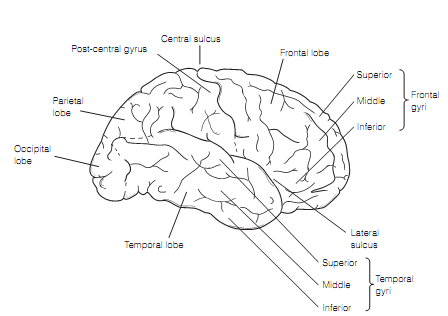
Figure: Lateral surface of the human right cerebral hemisphere.
The surface is covered by cortex and is highly complicated giving it a high surface area/volume ratio. The folds are known as gyri, and the creases between them sulci. Most of the cerebral cortex is neocortex that has six layers. The Cortical regions are mapped into Brodmann regions depends on the differences in cellular makeup and relative thickness of the layers. The importance of this is that the Brodmann map corresponds quite well to how functions are localized in the cortex.
The layers of the cerebral cortex are numbered from I, nearby to the pial surface through to VI that is the deepest as shown in figure. The layers contain various proportions of two types of neurons, the pyramidal cells that are output cells, and stellate cells which are interneurons. The relative thickness of the layers differs with the function of the cortical region. For illustration, the sensory cortex has a thick layer IV as of its large number of thalamic inputs, while motor cortex has a thick layer V as this is the position of motor neurons projecting to the brainstem and spinal cord.
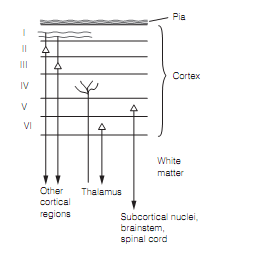
Figure: Inputs and outputs of the cerebral cortex.
The cerebral cortex is occupied in most of the brain activities, but is most frequently realted with the planning and execution of sensory perception, intentional movement, and cognitive functions. Those regions that are not particularly devoted to sensory or motor activities are known as the association cortex.
Within the core of each hemisphere lie clusters of nuclei which form the components of two neural systems, the extrapyramidal system and the limbic system as shown in figure. The major component of the extrapyramidal system, dependable for organizing stereo typed patterns of movement, is the basal ganglia, containing the striatum that lies in forebrain, and two midbrain nuclei, subthalamus and the substantia nigra. The striatum is partitioned into the neostriatum, itself composed of the two nuclei, caudate and putamen, and paleostriatum or globuspallidus. The putamen and globus pallidus altogether form the lentiform nucleus.
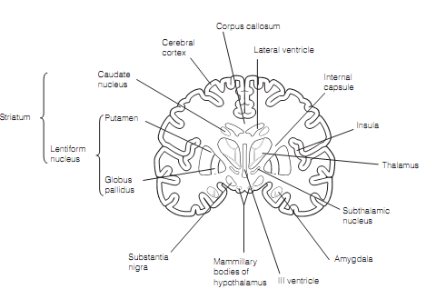
The limbic system comprises the nuclei (septal nucleus, amygdala, and mammillary bodies) plus areas of cerebral cortex that form a ring around the diencephalon, involving the cingulate cortex and hippocampus as shown in figure. The hippocampus is archaecortex and has only three layers. The hippocampus and amygdala are concerned with certain types of learning, and the limbic system in common is implicated in emotion.
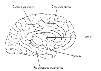
Figure: Medial surface of the human left cerebral hemisphere.