Numerical Aperture And Resolution:
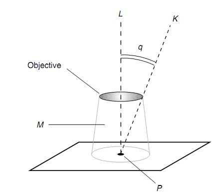
In an optical microscope, the numerical aperture of the objective is a significant specification in establishing the resolution, or the quantity of detail the microscope can provide. This is defined as shown in figure below.

Figure: Determination of the numerical aperture for a microscope objective.
Assume L be a line passing via a point P in the specimen to be inspected, and also via the center of the objective. Assume k be a line passing via P and intersecting the outer edge of the objective lens opening. (It is supposed that this external edge is circular.) Assume q is the measure of the angle among lines L and K. Assume M be the medium among the objective and the sample under inspection. This medium M is generally air, though not forever. Assume rm be the refractive index of M. Then the numerical aperture of the objective Ao is given by
Ao = rm sinθ
In common, the greater the value of Ao, the superior is the resolution. There are three manners to raise the Ao of a microscope objective of a specified focal length:
(A) The diameter of the objective can be raised.
(B) The value of rm can be raised.
(C) The wavelength of the enlightening light can be reduced.
The Large-diameter objectives containing short focal lengths, thus providing high magnification, are hard to build. Therefore, whenever scientists want to inspect an object in high detail, they can use blue light, that has a associatively short wavelength. Alternatively, or additionally, the medium M among the objective and the specimen can be modified to something with a high index of refraction, like clear oil. This shortens the wavelength of the enlightening beam which hits the objective since it slows down the speed of light in M. (Recall the relation among the speed of an electromagnetic disturbance, the wavelength, & the frequency!) A side effect of this tactic is a decrease in the effective magnification of the objective lens, though this can be remunerated for by using an objective with a lesser radius of surface curvature or by rising the distance among the objective and the eyepiece.
The use of monochromatic light instead of white light gives another benefit. The chromatic aberrations affect the light passing via a microscope in the similar way which it affects the light passing via a telescope. When the light has only one wavelength, chromatic aberration does not take place. Additionally, the use of different colors of monochromatic light (yellow, red, orange, green, or blue) can emphasize structural or anatomic features in a specimen which do not forever show up well in the white light.