Gaze stabilization
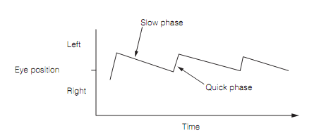
The head rotation detected by the semi-circular ducts triggers equivalent and opposite rotation of both eyes, a vestibulo-ocular reflex (VOR). For huge head rotations the eyes cannot continue to rotate but should be reset to a central position by speedily moving in the similar direction as the head. This gives mount to nystagmus, eye movements characterized by a slow phase which stabilize the retinal image, and quick phase which resets the eyes. By convention the direction of the nystagmus is the direction of the quick phase as shown in figure below.

Figure: Leftward nystagmus during the head rotation.
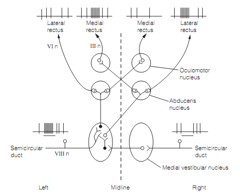
The horizontal semi-circular ducts are efficiently wired to the medial and lateral rectus muscles to generate the eye movements which counter the head rotation as shown in figure below.

Figure: The circuitry for vestibulo-ocular reflex. The stimulation of horizontal semi-circular ducts by the leftward rotation of the head excites the ipsilateral medial rectus and the contralateral lateral rectus and inhibits their antagonists. Excitatory neurons, open circles; and inhibitory neurons, filled circles. The Firing patterns are shown for cranial nerve neurons.
The gain of the VOR (i.e., the magnitude of the eye rotation divided by the magnitude of the head rotation) is pretty close to one for rapid head rotations. This means that there is a good match between eye and head movements that makes for a stable retinal image. The VOR can be altered by visual experience. Whenever human subjects wears magnifying lenses that means that they must make bigger eye movements to match head rotations, the profit of their vestibulo-ocular reflexes increases suitably over the next few days. The cerebellum is needed for this adaptation to take place, but not for it to be maintained once established. The instability of the retinal image proceeds as an error signal that is relayed to the cerebellum from the inferior olivary nucleus. The cerebellum studies to minimize the error and modifies its drive to the extraocular muscles. This is an illustration of motor learning. The lesions of vestibulocerebellum impair the capability to maintain steady gaze, causing unsuitable nystagmus.
The slow rotation of the head causes an apparent movement of the visual world in the reverse direction known as retinal slip; this is detected by the large, movement sensitive retinal ganglion cells and is used to produce eye movements that are equal in speed but in reverse direction to the retinal slip. This is the optokinetic reflex. As in the VOR, nystagmus takes place for large head rotations.