Extraocular eye muscle control
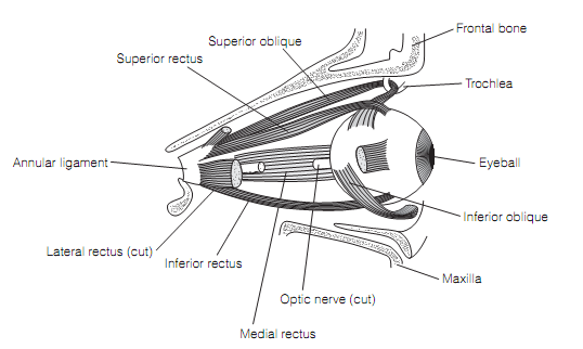
Each eye is moved by three pairs of extraocular eye muscles. The two pairs of rectus muscles (i.e., superior and inferior, medial and lateral) originate from a general annular tendon attached at the back of the orbit. Such muscles insert into the sclera in front of the equator of the eyeball. The third pair is the oblique muscles (i.e., superior and inferior) that insert into the sclera behind the equator of the eyeball as shown in figure below.

Figure: The right orbit displaying the extraocular muscles.
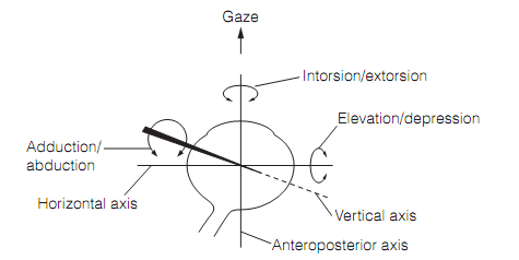
Working in concert these muscles proceed to rotate the eye about three principal axes as shown in figure below. The actions of the medial and lateral rectus muscles are very simple. They cause the eye to rotate about the vertical axis, therefore the gaze moves horizontally. The medial rectus brings rotation towards the midline (i.e., adduction) whereas the lateral rectus causes lateral rotation (i.e., abduction). The other two pairs of muscles generate rotations which have components along two of the principal axes, and the components modify depending on the horizontal location of the eye. These actions are concluded as shown in table below.

Figure: Principal axis for rotation of the eye, exposed for the right eye. In health, torsional movements (rotation around the anteroposterior axis) are small.
Eye muscles proceed in complementary fashion in the two eyes during conjugate movements in which the two visual axes move in parallel. Therefore, contraction of the lateral rectus in one eye is coupled with contraction of the medial rectus in the other eye for a conjugate horizontal shift in gaze as shown in table.

Table: Actions of extraocular eye muscles
The extraocular muscles are innervated by motor neurons in the nuclei of the oculomotor (III), trochlear (IV), or abducens (VI) cranial nerves. Such neurons are the final general path for the output of all five eye movement systems and are driven by brainstem reticular and medial vestibular nuclei axons which run in the medial longitudinal fasciculus. Eye motor neurons fire together statically, in a way relating to eye position, and dynamically, were reflecting eye velocity. To hold the eye steady in a given location needs tonic discharge by a specific set of motor neurons. The set will be distinct for various positions.
Eye movements are fetching about by pulses of action potentials with a firing frequency which is directly proportional to the velocity of the movement. This brings the eye to a new position that needs the generation of a new position signal. This is possibly achieved by integration of the velocity signal by the vestibulocerebellum and prepositus nucleus of the brainstem reticular system.