Noradrenergic pathways
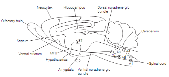
The Cell bodies of noradrenergic neurons are positioned in the pons and medulla (that is, cell groups A1–A6, except A3) as shown in figure. The main caudal groups A1 and A2 send their axons into the spinal cord where they form the synapses with terminals of primary afferents. The another project in two bundles, a dorsal and a ventral bundle, that unite to form the medial forebrain bundle (MFB) which ascends to bring the amygdala, hypothalamus, thalamus, hippocampus, limbic structures, and neocortex. The main noradrenergic cell group is the locus coeruleus (LC, group A6) that contributes most of the axons of the dorsal noradrenergic bundle and projects to the cerebellum. The Noradrenergic neurons are minute with fine, highly branched axons which ramify broadly. The axons bear varicosities along their length, but they do not form the close synaptic contacts, therefore noradrenaline (norepinephrine) is able to diffuse to reach the widespread targets. This is known as volume transmission. The Firing of noradrenergic cells is low in sleeping animals and rises with arousal level. Therefore noradrenaline (norepinephrine) is occupied in controlling sleep–waking cycles and in maintaining the arousal. It increases the signal-to-noise ratio of cortical procedure.

Figure: Major noradrenergic pathways in a sagittal part of the rat brain. A6 is the locus coeruleus. Here, MFB=medial forebrain bundle; ST=stria terminalis.