Peripheral auditory processing
In this part we will look at how the frequency selectivity found beside the basilar membrane is preserved or altered by the auditory nerve and how information about the intensity of the signal is encoded in the reaction of the auditory nerve fibres.

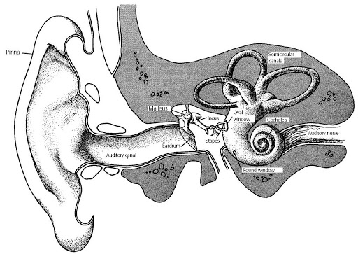
The nerve which communicates with or innervates the hair cells beside the basilar membrane is known as the vestibulocochlear nerve or VIIIth cranial nerve. It enters the brainstem merely under the cerebellum and conveys information from the hair cells in the inner ear and also from the vestibular organs of the inner ear. The cochlear section of the nerve (auditory nerve) contains two fundamental kinds of auditory nerve fibres: afferent fibres which carry information from the peripheral sense organ (i.e., organ of Corti) to the brain; and efferent fibres which bring information from the cerebral cortex to the periphery.
Afferent fibres occur from nerve cell bodies in the spiral (or cochlear) ganglion and contact the hair cells. The hair cells themselves do not contain axons and therefore do not produce action potentials. Action potentials are first generated by the axons of afferent fibres. Recall that approximately 10 per cent of the ion channels are open whenever the hair cell is unstimulated. This means that in the auditory nerve, there is a continuous low level of release of action potentials even when hair cells are unstimulated. The Depolarisation of hair cells in response to stereocilia shearing causes an increase in the discharge rate of action potentials above this spontaneous rate whereas hyperpolarisation of hair cells leads to reduce in the discharge rate of action potentials below the spontaneous discharge rate (inhibition).