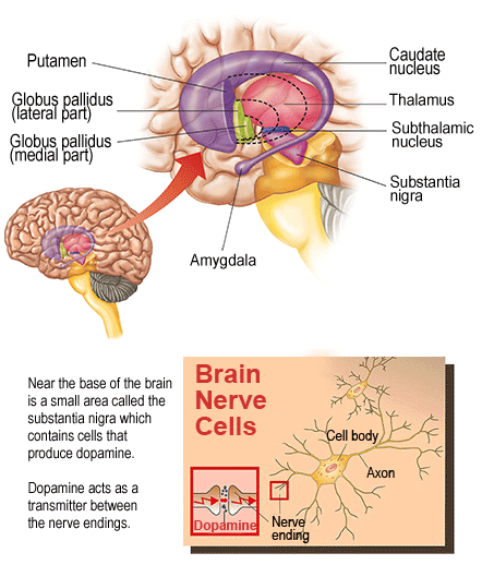
Anatomy of the basal ganglia
A collection of brain nuclei are collectively known as the basal ganglia. The basal ganglia which are studied in this section are the caudate nucleus, the putamen, subthalamic nucleus, the globus pallidus, and substantia nigra. The motor parts of the basal ganglia build up the extrapyramidal motor system, a word which is sometimes still used clinically.
A disease of the basal ganglia causes movement disorders. Movement disorders are diseases illustrated by hypokinesia) and/or hyperkinesia (abnormal involuntary movements like tremor and writhing movements). The Basal ganglia diseases involve such well-known diseases like Parkinson's disease (hypokinetic) and Huntington's disease (hyperkinetic). Though, not all movement disorders are due to the dysfunction of the basal ganglia.

An axial portion gives a panoramic view of the common shapes and relations of the caudate nucleus, & globus pallidus with each other and with the lateral ventricle and internal capsule. A coronal view via the frontal horn shows the associations of the head of the caudate and the anterior section of the putamen to the anterior limb of the internal capsule and the frontal horn. The putamen and caudate nucleus are developmentally a solitary nuclear mass which is divided by the internal capsule, though with gray matter bridges still connecting them. Since of this emergence, the putamen and caudate nucleus together are termed as the dorsal striatum. In Huntington's disease, the atrophy of striatum causes flattening of the caudate nucleus wherever it bulges into the lateral ventricle.
The putamen and globus pallidus altogether are termed as the lentiform nucleus. This name was given to them since of their lens shape as seen in axial portions.