Stimulated-emission depletion microscopy
Several super-resolution imaging techniques have been developed which allow light microscopy to obtain information far below the l/2 diffraction limit of visible light. One is stimulated-emission depletion microscopy (STED) which circumvents the diffraction limit by targeted de-excitation of dye molecules without making them fluoresce. Essentially this switches the dye molecules off, reducing the size of the excited region. STED works by using two lasers, an excitation laser and an offset laser. Fluorescent dyes which label specific sites of a sample are excited by light of a particular wavelength produced by the excitation laser. After a few nanoseconds the dye molecule spontaneously relaxes to the ground state by emitting a fluorescence photon of longer wavelength; that is, a redder light.
However, an excited molecule can also return to its ground state by stimulated emission instead of spontaneous relaxation and fluorescence. This can be triggered by irradiating the excited dye molecule with light of similar wavelength to the fluorescence light. This is done with the offset laser. In response, the dye molecule returns to the ground state, emitting a photon of exactly the same wavelength but without emitting a fluorescence photon. The offset laser light, stimulated emission, and fluorescence can be separated by appropriate optics. The offset laser is focused so as to de-excite (by stimulated emission) almost all of the dye molecules in a circular region activated by the excitation laser. The de-excitation zone is a broad annulus, leaving just a few fluorophores in the center of the region excited and hence able to fluoresce in the Figure. This allows nanoscale resolution (< 10 nm) permitting extremely fine detail to be visualized in neuron morphology that could hitherto only be approximated by three-dimensional reconstruction from ultra- thin (~50 nm) serial sections viewed under electron microscopy, a far more laborious technique. In addition STED can be done with live neural tissue, unlike electron micros- copy. This means that nanoscale changes in neuroanatomy can be followed over time.
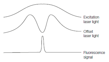
Figure: Stimulated emission depletion microscopy achieves super-resolution imaging by using an optical configuration that generates regions where fluorophores de-excite without fluorescing. Note the resemblance to surround inhibition