Confocal laser scanning microscopy
Conventional optical microscopy has a resolution limited approximately through half the wavelength of light. There are numerous kinds of light microscopy but the technique which gets closest to the limits imposed through the physics of light diffraction is confocal micros- copy. However, because it uses fluorescence to image a sample and fluorescent dye can be taken up by living cells; unlike other kinds of light microscopy fluorescence micros- copy can image living tissue.
A fluorescent dye molecule (photoactivatable fluorophore) is excited by light of a specific wavelength so that electrons are boosted from the ground state to high quantum energy states. After a few nanoseconds the molecule de-excites back to the ground state, emitting lower energy photons with longer wavelength because some of the energy of the exciting photon is dissipated internally within the molecule. In confocal microscopy the sample is illuminated by laser light tuned to excite the fluorescent dyes in the sample. However, rather than wide field illumination, as is the case with conventional light microscopy, light is brought to focus on a small region of the sample. In confocal laser scanning microscopy (Figure 1) scanning mirrors move the laser across the specimen so that each small region of it can be excited in turn. The fluorescent light emitted by the specimen passes through a pinhole before entering a digital camera. This eliminates all out of focus light; that is, only light from the focal plane reaches the camera.
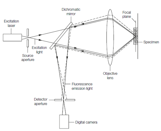
Figure: Principle of confocal microscopy. Excitation light is transmitted through a dichromatic mirror and brought to focus in the specimen. Emitted fluorescence light is reflected through a pinhole detector aperture into a digital camera. Fluorescence emitted from sources not on the focal plane (dashed lines) is cut off by the detector aperture.
The resolution of such a microscope is limited by the size to which the excited region can be focused and means that structures closer than 200 nm in the focal plane cannot be resolved. The depth resolution (along the optical axis) is worse, around 500 nm.