Action potential:
An electric potential which is known as the membrane potential exists across the plasma membrane of all cells. Many cells are electrically inactive as this membrane potential does not vary with time. Moreover, muscle and neurons cells are electrically active as their membrane potential can vary with time. In all cells the membrane potential is generated by the action of the Na+/K+-ATPase, with a high concentration of K+ inside the cell and a high concentration of Na+ outside. Neurons vary their electric potential through controlled modifications in the permeability of the plasma membrane to Na+ and K+ ions. Stimulation upon, the membrane potential of a neuron increases rapidly from the resting potential of -60 mV (millivolts) to around 40 mV shown in the figure; the membrane is said to depolarize and an action potential is generated. In order for this to happens, the membrane potential has to be depolarized beyond a critical threshold steps (around -40 mV). With time the membrane potential returns to the resting potential. An action potential is propagated along the axon beginning from the axon hillock.
The action potential arises from large, transient modifies in the permeability of the plasma membrane of the neuron to Na+ and K+ ions. Two kinds of voltage- sensitive ion channels are present in the membrane: one is selectively permeable to Na+ ions, the other to K+ions. These integral membrane proteins are sensitive to the membrane potential undergoing
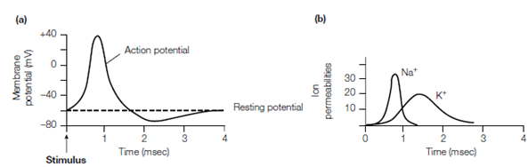
figure: The action potential. (a) Depolarization of the membrane potential; (b) changes in the permeability of the plasma membrane to Na+ and K+.
conformational modifies as the potential alters. first one is the conductance of the membrane to Na+ changes. The Depolarization of the membrane beyond the threshold steps causes a conformational change in the Na+ channel permits Na+ ions to flow down their concentration gradient from the outside of the cell into the interior which is shown in the figure. The entry of Na+ additionally depolarizes the membrane which causing more Na+ channels to open resulting in a rapid influx of Na+ and a modify in the membrane potential from -60 mV to 40 mV in a millisecond. The Na+ channels then spontaneously close and the K+ channels open permits K+ ions to flow out of the cell and restore the negative resting potential within a few milliseconds that is shown in above figure. The wave of depolarization is propagated along the axon
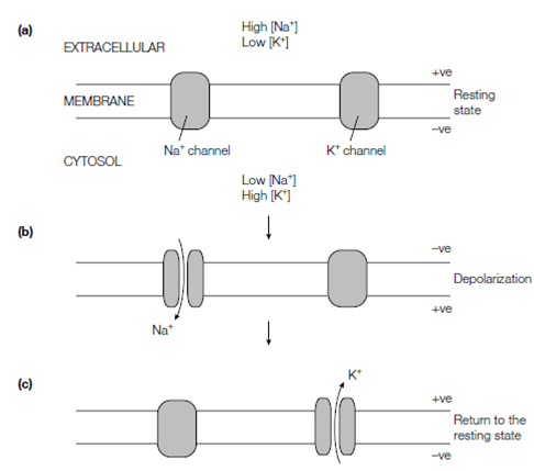
figure: Mechanism of depolarization of the nerve membrane by the opening and closing of selective Na+ and K+ ion channels.
through the opening of Na+ channels on the nerve terminal side of the initial depolarized region. The action potential can only move in that direction as the Na+ channels has a refractory period when they are insensitive to additionally stimulation. Only around one in a million of the Na+and K+ ions in a neuron flow across the plasma membrane during the action potential. Therefore, this is a very efficient pathway of signaling over long distances.
The tetrodotoxin, neurotoxin, a highly potent poison from the puffer fish, blocks the conduction of nerve impulses along axons and so leads to respiratory paralysis through binding very tightly to the Na+ channel and blocking its action.