Neuromuscular junction
The axon of a motor neuron splits into a number of branches at the surface of the muscle fiber. Each branch ends in a bouton that forms a synapse with the muscle fiber, known as neuromuscular junction (nmj). The cleft of the nmj as shown in figure is about 50 nm across. The postjunctional membrane, and the endplate that is thrown into folds, has a high density of nicotinic acetylcholine receptors under the active zones where acetylcholine (ACh) is discharged. The cleft consists of a collagenous basement membrane (basal lamina) to which is bound acetyl cholinesterase (AChE). The soluble forms of similar enzyme are also secreted into the cleft.
Nicotinic acetylcholine receptors (nAChR) are ligand-gated ion channels which mediate rapid ACh transmission. Binding of ACh opens the channel, permitting Na+ influx and K+ efflux. The reversal potential for this current is close to 0 mV, therefore activating nAChR causes depolarization.Spontaneous discharge of a single quantum of ACh at the nmj causes a 0.4 mV depolarization at the endplate known as miniature endplate potential (mepp). The arrival of an action potential at the motor nerve terminal triggers the discharge of 200–300 quanta generating numerous mepps that sum to generate a depolarization to around -20 mV, an endplate potential (epp). This greatly exceed the threshold for activating voltage-dependent sodium channels in the muscle membrane, therefore an action potential that is propagated over the muscle fiber membrane. The neuromuscular junction is exclusive among vertebrate synapses in that firing of the motor neuron almost invariably outcomes in the triggering of muscle action potentials.
The concentration of ACh reaches 1 mM in the nmj in around 200 µs of the arrival of an action potential at the motor nerve terminal though within a millisecond or therefore the ACh concentration has fallen back to baseline levels as of the high activity of AChE in the cleft. The enzyme hydrolyzes ACh to choline and acetate. The choline is taken back into the nerve terminal via a Na+ -dependent transporter.
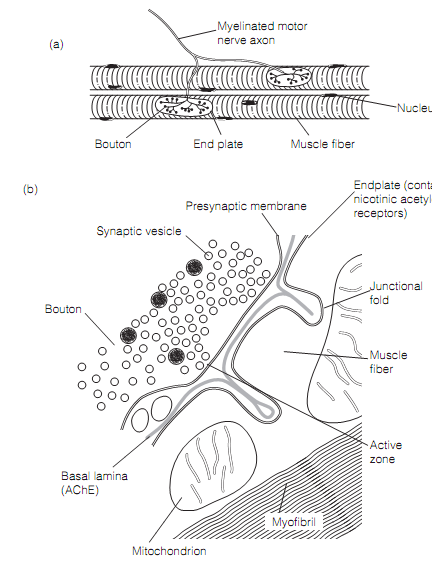
Figure: The neuromuscular junction: (a) a motor neuron forming synapses On two muscles fibers (x150); (b) a drawing of an electron micrograph of a neuromuscular junction.