Transmission and scanning electron microscopy
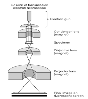
The Transmission electron microscopy utilizes electrons rather than photons to image specimens. This method was originally established from X-ray crystallography and it took some years to develop the method for biological specimens. In the electron microscope electron beams are focused through a series of electromagnetic lenses in Figure 7. The microscope is kept under high vacuum to insure unimpeded travel of electrons to and by the specimen. The electrons pass by the specimen on to a phosphor screen for visualization we cannot see electrons with our eyes. The Biological specimens are inherently unstable under vacuum and thus have to be fixed usually glutaraldehyde and paraformaldehyde followed through osmium tetroxide, dehydrated and stabilized in resins before they can be placed in a vacuum. The depth of specimen in which electrons can pass through is very small that means in which the biological specimen has to be sliced into thin sections usually with a diamond knife to produce 1 micron sections. These sections are electron transparent and specimen contrast has to be enhanced using metal stains example for, lead citrate and uranyl acetate. This method was used extensively for the study of subcellular structures and now has moved on to resolution at the molecular level. When the chemical fixation methods described above are replaced through cryo fixation and stabilization, tissue artifacts caused by traditional sample processing are reduced. The electron microscope can also be used to study organelle and virus structure using the method of negative staining. The Contrast is brought to the specimen through surrounding it with phosphotungstic acid.

Figure : Transmission electron microscope
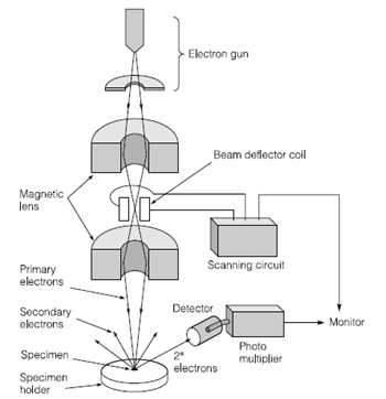
External structures of microbes can be viewed at high magnification using the method of scanning electron microscopy. The specimen is stabilized through either chemical or cryo fixation, covered through a layer of conductive material carbon or gold/platinum and the specimen is scanned with an electron beam. The Electrons are either reflected or passed by the specimen and a digital image is creating from the composite data, creating 3-dimensional structures in Figure 8. This technique has been further establishment for examination of specimens at low vacuum and ambient conditions.

Figure : Scanning electron microscope