Light microscopy
Of the organisms classed as microbes only the larger members of the chlorophyta and fungi are visible simply with the naked eye. A few members of the microbes are among 200 and 500 mm in size and can be seen using a hand lens but through far the largest group of microbes have cell sizes of among 1 and 10 mm. This last group needs substantial magnification before they can be viewed through the human eye and thus different types of microscope have to be used to see these microbes.
The first microscopes were based on simple, single lenses that provided sufficient magnification to see yeasts, protists and larger Bacteria. The early microscopists Robert Hooke in 1665 and Antonie van Leeuwenhoek in 1684 both observed and drew microbes using these simple single-lens microscopes. Present day light microscopes are compound microscopes they have an objective lens up to 100-fold magnification and an eyepiece lens usually 10-fold magnification in Figure 1 which together give a total magnification of 1000¥. At maximum magnification optical oil is used among the sample and the lens to optimize light collection and improve resolution in Figure 2. The resolution limit of the light microscope is about 0.2 mm example for you can differentiate two small black spots 0.2 mmapart limited through the physics of glass lenses. This level of magnification and resolution allows us to see most of the prokaryotes using a light microscope particularly if stains are used to maximize contrast among the background and the specimen.
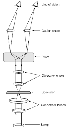
Figure 1. The light microscope
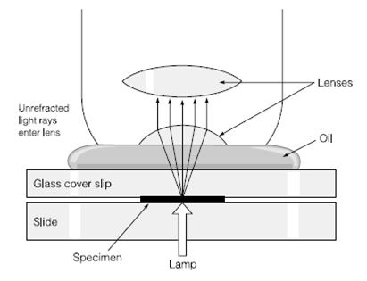
Figure 2. Effect of oil on the path of light through an objective lens
A number of stains can be used to visualize microbial cells for light microscopy including crystal violet and safranin that are both used in the Gram stain. The Bacterial spores can be stained using malachite green and fungal structures can be stained using cotton blue in lactophenol. All stains should be treated with great care as through their very nature they are toxic to cells including yours.
Other microscopic techniques can be used to maximize contrast between specimen and background including phase fluorescence, contrast and dark field microscopy. The Phase contrast microscopy takes advantage of the change in phase of light which occurs when light passes by a cell. This change in phase alters the refractive index of the sample relative to the background and when this difference is amplified using a phase plate in the microscope the resulting image has enhanced contrast in Figure 3. Dark field micros- copy uses side illumination of the specimen. Only scattered light from the specimen is seen through the microscope in Figure 4 and the object appears light on a dark back- ground. Fluorescence microscopy takes benefits of the fact in which some molecules will emit light fluoresce when irradiated with light of another wavelength. Some microbes contain naturally fluorescent compounds chlorophylls and other pigments. Fluorescent dyes can be used to tag specific structures or processes. Light from the ultraviolet spectrum is commonly used to excite fluorescence and this requires a separate source of illumination.