Anatomy of learning
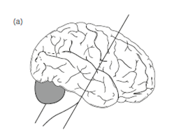
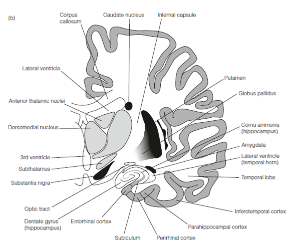
Considerable evidence implicates the medial temporal lobe in the given Figure and its connections as critical for declarative learning. Bilateral lesions of the hippocampus even when confined to the selective loss of pyramidal cells from just one region result in anterograde amnesia so extreme in which patients are no longer able to form new long-term memories. WM and procedural memory are unharmed as are remote long-term memories, while some retrograde amnesia is seen.
Bilateral medial temporal lobe lesions in macaque monkeys give an animal model for human amnesias. Usually, the animals are trained on a delayed nonmatching to template task. In this case the monkey is trained to select an object to obtain a food reward. Following a variable delay during in that the animal cannot see any manipulations, it is given a choice among the similar object and a novel object and is needed to select the novel object to get the reward, which is, it required to remember that object it saw first. Lesioned animals show anterograde amnesia and selective lesioning shows that the most severe deficit occurs with damage to the parahippocampal and perirhinal cortex of the temporal lobe.
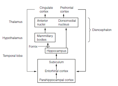
Three diencephalon structures are extensively connected with the temporal lobe and play a function in memory. The major output of the hippocampus is the fornix that projects largely to the mammillary bodies of the hypothalamus and output from that goes to the anterior thalamus.


Figure: Section (a) through the human brain to show (b) the gross anatomy of the medial temporal lobe.
In addition, areas of the amygdala and temporal cortex make connections of the thalamus with the dorsomedial nucleus. Bilateral lesions confined to just one of these diencephalonic structures in monkeys modestly impairs performance in the delayed nonmatching to samples tasks but larger lesions affecting all three produce very severe deficits. The proposed circuitry for declarative memory is described in Figure.

Figure: Mammalian forebrain memory circuitry.