Phosphoinositide second messenger system
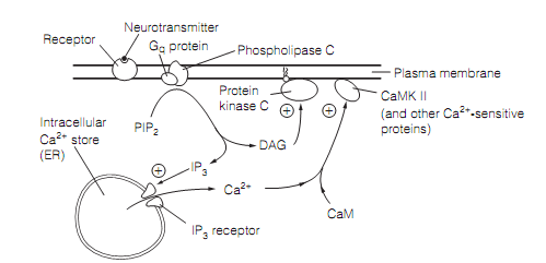
Most of the receptors are coupled through the Gq protein for the activation of phospholipase C which is as shown in figure. This enzyme cleaves a small phospholipid in the inner leaflet of the plasma membrane, that is phosphatidyl inositol-4, 5-bisphosphate (PIP2), to provide diacylglycerol (DAG) and inositol-1, 4, 5-trisphosphate (IP3), together of which are the second messengers. The DAG, a hydrophobic molecule, diffuses in the lipid where it activates the protein kinase C (PKC). In return this kinase phosphorylates its protein targets, affecting the receptor, metabolic, and ion channel functions.

Figure: A diagram of phosphoinositide second messenger is shown. Here CaM=calmodulin; CaMKII=calcium-calmodulin-dependent protein kinase II; DAG=diacylglycerol; ER=endoplasmic reticulum; IP3=inositol trisphosphate; PIP2=phosphatidyl inositol bisphosphate.
The IP3 is water soluble and freely diffusible in the cytosol. Its aim is the IP3 receptor, a big IP3-gated calcium channel situated in the membrane of smooth endoplasmic reticulum (SER). The SER in neurons (and its corresponding, the sarcoplasmic reticulum in muscle cells) acts as an intracellular Ca2+ store. The binding of IP3 to its receptors causes the calcium channels to open and Ca2+ flows out of the SER into the cytosol. The rise in intracellular calcium concentration has diverse and widespread effects which are cell usual. An obvious illustration is that by binding the protein troponin in a striated muscle, calcium triggers cascade of the biochemical events which leads to the muscle contraction. The Neurons holds calcium binding protein known as calmodulin (CaM) that shares considerable homology with troponin. On binding Ca2+, the calmodulin activates a number of enzymes involving calcium–calmodulin-dependent protein kinase II (CaMKII). The CaMKII, and many other calcium sensitive proteins, mediate the effects of the raised intracellular calcium, like the changes in the gene expression and membrane permeability.