Fungal wall structure and growth
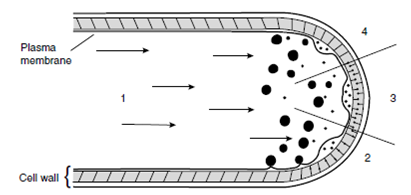
Fungal walls are rigid structures formed from layers of semi-crystalline chitin microfibrils that are embedded in an amorphous matrix of b 1–3 and b 1–6 glucan. Some protein may also be present. Growth occurs at the hyphal tip by the fusion of membrane-bound vesicles containing wall-softening enzymes, cell wall monomers, and cell wall polymerizing enzymes derived from the Golgi with the hyphal tip membrane. The fungal wall is softened extended through turgor pressure and then rigidified.

Figure : Hyphal tip growth. Step 1, vesicles migrate to the apical regions of the hyphae. Step 2, wall-lysing enzymes break fibrils in the existing wall, and turgor pressure causes the wall to expand. Step 3, amorphous wall polymers and precursors pass through the fibrillar layer. Step 4, wall-synthesizing enzymes rebuild the wall fibrils. From Isaac S (1991) Fungal– plant interactions. With permission from Kluwer Academic.
Septae are cross walls that form within the mycelium. Growth in the Zygomycota and Chytridiomycota is not accompanied by septum formation, and the mycelium is coenocytic. Septae only occur in these groups to delimit reproductive structures from the parent mycelium, and they are complete. In the dikarya growth of the mycelium is accompanied by the formation of incomplete septae. Septae in the Ascomycota are perforate, and covered by ER membranes to limit movement of large organelles such as nuclei from compartment to compartment. This structure is called the dolipore septum. In dikaryotic Basidiomycota, septum formation is coordinated with divisions of the two mating-type nuclei, maintaining the dikaryotic state by the formation of clamp connections. These septae resemble crozier formation in the formation of asci and the structures may be homologous.