Structure of the eye
The three layers surround the contents of the eye, the sclera, the choroid and the retina as shown in figure. The sclera is a stiff, thick, outer layer of connective tissue. At the front of the eye it becomes the cornea. At the back it becomes the dura mater covering the optic nerve. The sclera preserves the shape of the eyeball and gives attachment for the extraocular muscles. The curvature of the transparent cornea refracts incoming light and gives most of the focusing power of the eye. The choroid is a highly vascular layer, thin, dark brown in color as of the presence of choroidal pigment cells. By absorbing light it restricts the total internal reflection within the eye.
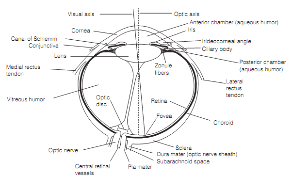
Figure: Horizontal part through the right human eye.
At the front the choroid becomes the ciliary body and the iris. The ciliary body provides rise to several, thin zonular fibers that attach to the capsule of the lens as the suspensory ligament. Inside the ciliary body lies the ciliary muscle which composed of smooth muscle fibers arranged in both the radial and circular directions.
The iris is a diaphragm enclosing a central hole, the pupil. The iris consists of two intra-ocular muscles that act in concert to control the size of the pupil. The Innermost is a flat ring of circularly arranged smooth muscle fibers, the pupillary sphincter. Enclosing the sphincter is a layer of radially organized myoepithelial cells that form the pupillary dilator.
The aqueous humor is actively secreted by the epithelium of the ciliary body into the posterior chamber. It percolates, however the pupil to the anterior chamber from where it drains into the venous system through the canals of Schlemm positioned in the irideocorneal angle. The Aqueous humor supplies the metabolic substrates for the lens and cornea thathave no blood supply, and maintains the eyeball pressure. The Vitreous humor is a gel of extracellular fluid that contributes to the refractive power. The biconvex lens of the human eye has a diameter of 9 mm. It is encapsulated in an elastic connective tissue membrane that is attached to the suspensory ligament.