Muscle spindle reflexes
The main elementary modulation of motor unit output is formed by sensory input from the muscle spindles that measure the length and rate of change of length (i.e., velocity) of the muscle. Any attempt to stretch the muscle rapidly, for illustration by rapidly loading it, is met by contraction. This is the muscle spindle reflex (i.e., stretch reflex and myotatic reflex) and is a negative feedback method that defends a constant muscle length in the face of external forces that act to perturb it. A stretch reflex can be extracted from any skeletal muscle by sharply tapping its tendon. The resulting stretch causes the muscle to contract. The stretch reflex is most easily elaborated by tapping the patellar ligament among its insertion into the kneecap and the tibia, causing the contraction of the quadriceps femoris, the powerful class of extensor muscles on the front of the thigh as shown in figure below.
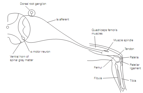
Figure: Basic circuit of a stretch reflex. Striking the patellar ligament excites some hundred la afferents.
The sensory side of stretch reflex consists of the muscle spindle and its afferents. Muscle spindles lay in parallel with the standard extrafusal fibers therefore any force acting on entire muscle acts in the similar way on spindle. Each muscle spindle is a fluid-filled capsule of connective tissue, 4–10 mm long and 100 µm in diameter, having about seven modified muscle fibers known as intrafusal fibers as shown in figure below. Intrafusal fibers contain contractile ends though their central areas are non-contractile. There are two kinds of intrafusal fiber, nuclear chain and nuclear bag.
Nuclear bag fibers (b) are swollen at their center, wherever their nuclei are clustered, and are innervated by the large diameter myelinated (Ia) primary afferents, the ends of that spiral around the central area of the fiber. There are two sorts of nuclear fibers bag that can be renowned by whether in addition to primary afferent innervation they also receive secondary, group II, myelinated afferents. Those which are not dynamic (b1), those which are static (b2).
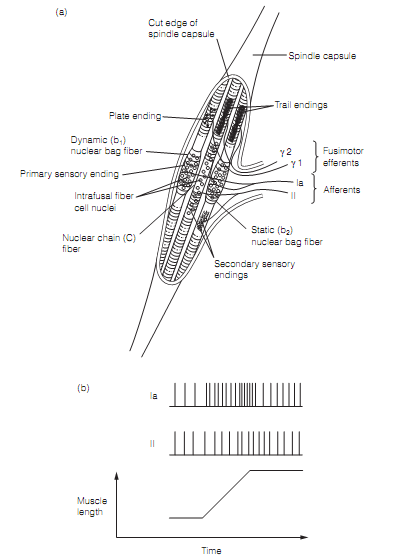
Figure: Muscle spindles: (a) a spindle opened to show intrafusal fibers and their innervations. A spindle generally contains one b1, one b2, and numerous c fibers; (b) responses of la and II afferents to muscle stretch.
Primary afferents explain dynamic responses; respond to the rate of change of length (i.e., velocity). This is as of the properties of the dynamic nuclear bag fiber. Whenever stretched the central area extends causing the Ia afferent to fire a volley of action potentials. Consequently, though, the poles of the fiber extend slowly allowing the central area to creep back to a shorter length therefore the firing rate of the Ia afferent drops off. Prime afferents also show signaling muscle length, static responses, by virtue of their innervation of static (b2) nuclear bag fibers that are stiffer than the dynamic fibers and therefore elongate in proportion to muscle stretch.
Nuclear chain fibers (c) are of regular diameter, are around half the size of b fibers and their central area contains a line of nuclei. They are innervated by primary and secondary afferents and being stiff (similar to b2 fibers) these afferents respond to muscle length. Usually a spindle will contain one b1, & b2, and 3-5 c intrafusal fibers.
The bulk of Ia spinal afferents form synapses on homonymous motor neurons, which is, motor neurons going to the same muscle. Though, about 40% make synapses with motor neurons that go to synergistic muscles. For illustration, the quadriceps femoris consists of four muscles which act synergistically. Afferents from spindles in any one of them will set up connections with its own motor pool and the pools of the other three muscles.
A stretch reflex has two components. The phasic component is that viewed by tapping the tendon of a muscle. It takes place rapidly, is short, and occurs since of the dynamic activity of the Ia afferents. The tonic component is much more constant contraction brought about by the static activity of the Ia afferents and the secondary, group II afferents. This component is mainly significant in keeping posture. Standing in a moving vehicle, for illustration, the muscles in the legs and trunk which are stretched by the swaying will be contracted, therefore keeping the body upright. A rapid jolt will, of course, also trigger the phasic component.
Cell size is bimodally dispersed in motor pools. The neurons that drive the extrafusal fibers to generate muscle contraction are Aα class with a cell body diameter averaging 80 µm, generally termed as motor neurons. Additionally there is a population of smaller cells belong to the Aγ class, known as γ motor neurons, axons of that (fusimotor fibers) go to the muscle spindles. All intrafusal fibers comprise their contractile ends innervated by these γ motor neurons. Contraction of the ends of intrafusal fibers maintains the central area taut therefore it can respond to muscle stretch. Therefore, one purpose of γ efferent discharge is to sustain that the sensitivity of the muscle spindle to alters in length over a broad range of lengths. Without it, the muscle contraction would cause the intrafusal fibers to slacken and fail to respond to stretch.
There are two class of γ motor neuron which can be activated separately by the CNS; γ1 (dynamic) innervate b1 whereas γ2 (static) innervate b2 and c fibers. Stimulation of γ1 fibers raises the sensitivity of the b1 fibers; therefore the primary afferent firing rate is very high in rapid than slow stretch. Stimulation of γ2 fibers enhances firing of secondary afferents in response to steady stretch. In both situations the γ efferents are increasing the gain of the spindle. Firing rates of γ efferents are raised whenever performing movements which are mainly complex.
Stretch reflexes should be overridden to permit the execution of a movement as the muscle should contract isotonically and shorten. This is accomplished by descending motor pathways exciting both α and γ motor neurons at similar time. This is known as coactivation. It makes the intrafusal and extrafusal fibers shorten altogether in such a manner that the intrafusal fibers are always adequately taut to respond to stretch.