Inverse myotatic reflex
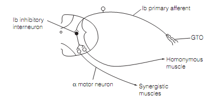
Positioned in the tendons, in sequence with muscle fibers are Golgi tendon organs (GTOs) that measure muscle tension. Increases in muscle tension start a negative feedback reflex, the inverse myotatic (i.e., Golgi tendon) reflex that opposes the increases in tension. It is brought around by GTO input activating inhibitory interneurons which synapse with motor neurons supplying the muscle as shown in figure.

Figure: Circuitry of the inverse myotatic reflex. An increase in muscle tension causes Golgi tendon organ (GTO) lb afferent to fire at a greater rate.
The GTO is composed of the collagen fibers that join muscle fibers to tendons, interwoven via which are axon branches of a group Ib afferent neuron. The increased tension which occurs with muscle contraction stretches the collagen fibers; distort the terminals of the Ib afferent that fires. Individual Ib afferents respond statically, reflecting the level of tension in response to the activation of a single motor unit. GTOs do not measure average tension of the muscle as less than one percent of fibers in motor units are coupled to GTOs.
The Ib afferents enter the spinal cord to synapse in the intermediary zone (Rexed laminae VI–VIII) on inhibitory neurons, which then synapses with motor neurons of the homonymous and synergistic muscles. Such inhibitory neurons are all particular to this disynaptic reflex pathway as shown in figure and therefore are designated Ib inhibitory neurons (IbINs). Inhibition of homonymous and synergistic muscle by GTOs is augmented by inputs from Ia spindle afferents, joint afferents, & cutaneous mechanoreceptor afferents onto IbINs. The functional significance of these connections is not clear. Descending motor systems might either excite or inhibit IbINs.