Lateral geniculate nucleus
The pathways for visual perception begin with the retinogeniculate fibers, the axons of ganglion cells which end in the lateral geniculate nucleus (LGN). Since of the way in which the fibers are sorted in the optic chiasm, the left optic tract and left LGN carry axons from the left side of both retinas. Therefore the left LGN shows the right side of the visual field.
The primate LGN has six layers which are as shown in figure. The two most ventral are the magnocellular layers that receive input from M ganglion cells and are concerned with motion. The Dorsal to these are four parvocellular layers which are innervated by the P ganglion cells and encode form and color. The interleaved between these major layers are koniocellular layers having small K cells. These receive input from the K ganglion cells and generally have small receptive fields. There are numerous classes of K cells. A few get input from the blue-on K ganglion cells and relay to the primary visual cortex (V1). The others are motion sensitive and bypass V1, projecting to the medial temporal cortex that is also the main target for M cells conveying the motion information.
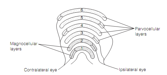
Figure: The structure and inputs of the lateral geniculate nucleus.
The LGN cells have circular RFs with enclose antagonism. They represent little or no response to the diffuse light covering the entire receptive field. Each layer in LGN gets input only from one eye and no cells show the binocular responses (i.e., responses to both eyes). It is not until the visual cortex is reached that input from both the eyes is combined. The responses of the LGN cells match those of the ganglion cells that supply them, therefore on and off channels remain independent and P cells display accurately the same color opponency properties as retinal ganglion cells. There is a very accurate topographic (retinotopic) mapping in ideal register onto each of the layers of the LGN, with the fovea taking up about half of the space.
The responses of LGN cells are smaller and can be elucidated by classic center–surround processing. The responses are suppressed by the retina and probably by inhibitory interneurons in the LGN itself. This suppression might act to permit the wide variety of contrasts seen in the natural world to be accommodated by the limited dynamic variety of neurons.
The LGN output is through geniculostriate neurons which project to the primary visual cortex (V1). Only around 10% of the synapses on these are from retinal ganglion cells. The other synapses are made by back projections from V1, the better colliculus, the parabrachial nucleus, and the thalamic reticular nucleus. These are probably included in visual attention; that is, in selecting which retinal input is transmitted through the visual cortex.