Dopaminergic pathways
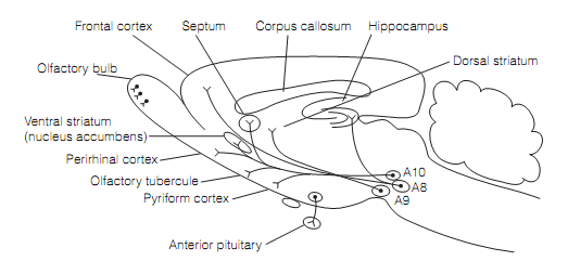
The Dopamine neurons are broadly distributed in the nervous system, being found in the olfactory bulb, retinal amacrine cells, and autonomic ganglia. Most, though, are confined to a few nuclei in the brainstem, sending their axons to many areas of the forebrain involving the cerebral cortex. These main pathways are illustrated in figure which is shown below.

Figure: Major dopamine pathways in a sagittal part of the rat brain. The A8 & A10 collection of dopamine neurons provide rise to the mesolimbic and mesocortical tracts. The nigrostriatal tracts originate in the substantia nigra (A9). The A12 neuron axons run in the tuberoinfundibular pathway.
Around 80% of the dopamine neurons are in zona compacta of the substantia nigra (SNpc) that constitutes the A9 group of catecholaminergic cells. (Such groups range from A1 to A16, the maximum the number the more rostrally they are positioned.) The SNpc neurons project to the striatum as the nigrostriatal pathway. Such cells are included in basal ganglia regulation of movement and their loss outcomes in Parkinson’s disease. The Dopamine cell clusters (groups A8 and A10) in the ventral tegmentum of the midbrain project to limbic structures (example, nucleus accumbens) or to related cortical regions (example, medial prefrontal and cingulate cortex), give rise to the mesolimbic and mesocortical systems correspondingly. These are implicated in motivation, drug addiction, and in the schizophrenia. Numerous small groups of dopaminergic cells in the hypothalamus project axons in the tuberoinfundibular pathway to the pituitary to inhibit the secretion of prolactin or growth hormone.
The Dopaminergic neurons are very small with a thin unmyelinated axon (that arises from one of the dendrites) that bears numerous varicosities along its length. The Action potentials in dopamine neurons are long lasting (2–5 ms) and propagated very slowly (0.5 m s-1).