DNA double helix:
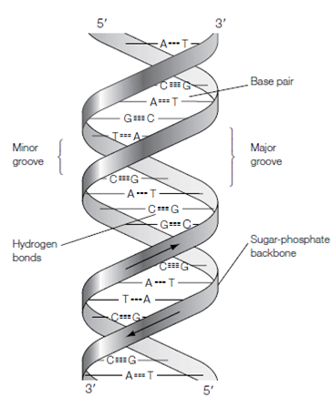
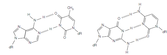
In the year of 1953, Watson and Crick worked out the 3-D structure of DNA, beginning from X-ray diffraction photographs taken through Wilkins and Franklin. They deduced which DNA is composed of two strands wound round every other to form a double helix with the bases on the inside and the sugar-phosphate back- bones on the outside. In the double helix that is shown in the figure, the two DNA strands are organized in an antiparallel arrangement example for the two strands run in opposite directions one is orientated 3'→5' and the other strand is orientated 5'→3'. The bases of the two strands form hydrogen bonds to every other; the pairs with T and G pairs with C. This is known as complementary base pairing. Therefore a large two-ringed purine is paired with a smaller single-ringed pyrimidine and the two bases fit neatly in the gap among the sugar-phosphate strands and maintains the right spacing. There would be insufficient space for two large purines to pair and too much space for two pyrimidines to pair that would be too far apart to bond. The A:T and G:C base pairing also maximizes the number of effective hydrogen bonds which can form among the bases; there are three hydrogen bonds among every G:C base pair and two hydrogen bonds among each A:T base pair. Therefore A: T and G: C base pairs form the most stable conformation both from steric considerations and from the point of view of maximizing hydrogen bond formation.

figure: The DNA double helix.

Adenine : thymine Guanine : cytosine
figure: The DNA base pairs. Hydrogen bonds are shown as dashed lines. dR, deoxyribose.