Transilluminator:
It is an ultraviolet light source that is used to visualize ethidium bromide-stained DNA in gels. After DNA gel electrophoresis is run, a whole slab is kept over transilluminator. The spots of DNA in the agarose slab can simply be found through the fluorescence of intercalated ethidium bromide.

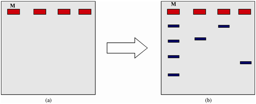
Figure: Slab electrophoresis separating DNA samples. Rectangle boxes are the wells generated by comb in agarose slab. (a) and (b) represent the gel-slab before and after run, respectively. Well M contains standard marker to examine the mass of the DNA.
Between linear and supercoiled DNA, the last will move faster in agarose slab as compared to the former since of the difference of surface area. Therefore, a plasmid DNA could simply be examined whether it is linear or supercoiled or a mixture of two. Actually, it is simple understandable which a nicked plasmid will move slower in DNA gel than the native one.
Pulse-Field Gel Electrophoresis (PFGE) permits investigators to separate much larger pieces of DNA than conventional agarose gel electrophoresis. Within conventional gels, a current is applied in a single direction (from top to bottom). Thus, in PFGE, the direction of the current is altered at regular intervals. Initially, the agarose gel is prepared and samples are loaded within the wells after mixing along with blue loading buffer. Then the current is turned on and the direction of the current is changed within a regular pattern. This is repeated until the loading dye reaches near the end of the gel.
The gel is then soaked within a solution holding ethidium bromide that fluoresces orange while bound to DNA.