Lateral motor pathways
There are two lateral pathways for downward control of voluntary movement. Both invent in the motor cortex that lies on the frontal lobe mere anterior to the central sulcus. The corticospinal tract consists of axons of around one million pyramidal cells in layer V of the cortex. More than half come from the main motor cortex (M1, Brodmann region 4) or secondary motor region (MII, Brodmann region 6). These axons project to the ventral horns of the spinal cord to modify the activity of α and γ motor neurons. Around 40% of corticospinal tract axons come from the somato-sensory cortex (Brodmann regions 1, 2, & 3) or other areas of the parietal cortex (Brodmann regions 5 &7). These axons terminate in the dorsal horns of the spinal cord and regulate the sensory input.
Most of the corticospinal tract consists of fine myelinated & unmyelinated axons with conduction velocities between 1 to 25 ms-1. Though, there are around 30 000 extremely large (20–80 µm diameter) pyramidal cells in region 4, known as Betz cells, with big myelinated axons which conduct with velocities of 60–120 ms-1. Axons of the corticospinal tract pack strongly to pass through the internal capsule that lies between the thalamus and the lentiform nucleus as shown in figure and descend into the brainstem. At this point the most medial fibers peel off and cross the midline to go to nuclei (trigeminal (V), facial (VII), and hypoglossal (XII), & accessory (XI)) of the cranial nerves. These are corticonuclear fibers and are motor to the tongue, face, pharynx, larynx, and sternomastoid and trapezius muscles. The residual axons descend via the medulla causing a swelling on its ventral surface, the pyramid, therefore the corticospinal tract is also known as the pyramidal tract. At the caudal medullas 85% of fibers cross the midline as the pyramidal decussation, providing rise to the lateral corticospinal tract. The residual ipsilateral axons form the anterior corticospinal tract that crosses over at spinal cord level.
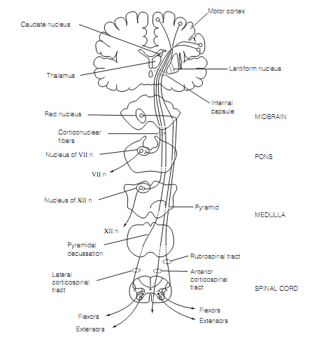
Figure: The lateral motor pathways. Only the corticonuclear fibers in the facial (VII n) and hypoglossal (XII n) nerves are shown. Synapses of the anterior (ipsilateral) corticospinal tract with spinal cord interneurons are not shown.
The Corticospinal tract neurons use glutamate as a transmitter and are excitatory. They either synapse directly with motor neurons delivering distal limb muscles in Rexed lamina IX, or synapse with interneurons in the laminae VII and VIII that make polysynaptic connections with motor neurons of axial muscles and proximal limb muscles. Fusimotor (γ-efferent) neurons that should be coactivated with motor neurons to override the stretch reflex during voluntary movement are excited polysynaptically. Stimulation of corticospinal tract is principally excitatory to flexors but inhibitory to extensors. The corticospinal tracts inhibit motor neurons disynaptically through Ia inhibitory interneurons.
The corticospinal tract axons occuring from the somatosensory cortex project to cranial nerve sensory nuclei and dorsal horns and generate presynaptic inhibition on main afferent terminals except for 1a spindle afferents. Few pyramidal cells in layer V of the motor cortex send their axons in the corticorubral tract to the red nucleus in the midbrain that also receives collaterals from the corticospinal tract. The red nucleus offer rise to the rubrospinal tract, the second of the lateral motor pathways. Few of its axons go to cranial nerve nuclei in the pons and medulla. In humans the rubrospinal axons descend as portion of the corticospinal tract.