Control of breathing
The diaphragm and muscles of the chest wall used in breathing are skeletal muscles supplied by motor neurons of the somatic nervous system. In spite of this we only have voluntary control over breathing (through pyramidal tract control of spinal motor neurons) for short time period as this is overridden by brainstem circuits which drive and regulate breathing. Such circuits get sensory input from visceral afferents—for illustration pharynx, airways baroreceptors, and chemoreceptors—and are inter-connected with CNS circuits controlling the cardiovascular system and sleep. This permits breathing to be altered in speech, swallowing, exercise, and sleep. Inputs from the limbic system evoke modifications in breathing due to emotions.
Breathing outcomes from the rhythmic relaese of spinal motor neurons that supply ventilatory muscles. The axons of motor neurons in spinal segments C3–C5 run in the phrenic nerves to the diaphragm, contraction of that raises chest volume during inspiration. The motor neurons in C4–L3 supply neck muscles and external intercostal muscles which aid inspiration, internal intercostal muscles and abdominal muscles answerable for expiration.
The spinal motor neurons are driven by a central pattern generator which consists of heavily inter-connected neurons in the medulla and pons as shown in figure below. The dorsal respiratory group (DRG) is clusters of neurons in the nucleus of the solitary tract (NST) of the medulla. One cluster is inspiratory upper motor neurons that synapse with spinal motor neurons supplying the diaphragm and external intercostal muscles. Such cells are active during quiet inspiration. They obtain inputs from another cluster of DRG cells which integrate the visceral inputs. The ventral respiratory group (VRG), in the ventrolateral medulla, is silent in quiet breathing though active in heavy breathing. The VRG houses both inspiratory and expiratory neurons that are reciprocally innervated by inhibitory connections. Therefore, when the inspiratory neurons are firing the expiratory neurons are silent and vice versa. The pontine respiratory group (PRG) involves the Kölliker–Fuse nucleus (KFN), that drives the medullary respiratory neurons therefore as to prolong inspiration (apneusis), and cells in the parabrachial nucleus (PBN) of the rostral pons that overrides apneusis, thereby terminating the inspiration.
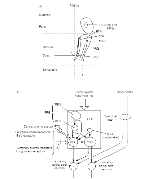
The fundamental respiratory rhythm could originate from the pre-Botzinger complex (pBOT), a nucleus just rostral to the VRG that makes extensive connections with the VRG, DRG, and pons. The Pre-Botzinger complex cells have inherent pacemaker properties. Integrating neurons in the DRG obtain sensory input, largely through the vagus nerve, from a variety of sources that alter the fundamental rhythm of breathing. These consist of the following:
? Nociceptors (lung irritant receptors) that trigger the cough reflex.
? Pulmonary stretch receptors that restrain inspiration.
? Baroreceptors, that inhibit inspiration: when blood pressure falls (as a outcome of hemorrhage, for illustration) depth of inspiration increases.
? Peripheral chemo receptors in carotid body and aortic arch that are activated by large deductions in partial pressure of O2.
? Central chemoreceptors that are excited by a increase in brain extracellular fluid concentrations of CO2 and fall in pH and drive deeper breathing in reaction. Central chemoreceptors have been specified in the retrotrapezoid nucleus that is adjacent to the nucleus of the facial (VII) nerve.