Receptive fields
The spatial position of a stimulus on a sensory surface (i.e., skin, retina, etc.) is given by that specific subset of neurons react. The receptive field (RF) of a neuron is the area of a sensory surface that when stimulated causes a change in the firing rate of the neuron. The Primary afferents normally have small RFs, the size of which is govern by the allocation of the cluster of sensory receptors that supply the afferent. The Receptive fields of neighboring neurons responding to similar kind of stimulus tend to overlap.
Further proximal neurons in a sensory pathway have receptive fields which are composites of the RFs of more distal neurons. This gives increase to two features which is explained below:
- In common, the proximal neurons have larger RFs since of convergence; numerous afferents might synapse on a single more proximal (that is, downstream) neuron. The Low convergence is seen where the high spatial resolution (that is the capability to sense stimuli which are close altogether as independent) is significant, such as among cones and bipolar cells in the retina. In contrary, the high convergence is needed where it is essential to integrate weak signals from a number of receptors to attain high sensitivity. This is the situation among rods and bipolar cells in the retina, where it allows the vision in dim light.
- The more proximal a neuron is the more complex is its receptive field. This is as the proximal neurons get inputs from a broader range of sources than distal neurons. This reflects the reality that extensive information processing takes place in sensory systems. The Greater complexity of receptive fields also increases as an effect of an extremely general characteristic of sensory pathways, lateral inhibition. At its easiest, this is where the RF of a neuron has two zones, the central area and a surround, from which the opposite and antagonistic effects are generated in the cell whenever stimulated. It is seen in somatosensory, visual, and auditory pathways. Figure shows the receptive field of a somatosensory cell. The Stimulation of the center causes a rise in firing therefore the RF is said to have an excitatory center. The Stimulation of the surround creases firing and is brought about by inhibitory interactions. The cell behaving in this manner is explained as an on-center cell. Off-center cells are also very general. For the on-center cell, maximum firing rate would be seen with a stimulus that merely managed to stimulate the whole center. A larger stimulus which encroached upon the surround would be less effective, by causing few inhibitions. In this manner the lateral inhibition sharpens spatial resolution and enhances the contrast at boundaries among stimuli.
In skin mechanoreceptor afferents these improve two-point discrimination. By the same means, light–dark contrast at edges is enhanced in the retina, and tone discrimination sharpened by the central auditory neurons. In common, lateral inhibition occurs between neurons coding similar kind of sensation. Though, color vision depends on lateral inhibition among cells which respond to various wavelengths.
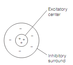
Figure: The Receptive field of an on-center sensory neuron displaying lateral inhibition; an off-center neuron would have an inhibitory center and an excitatory surround.
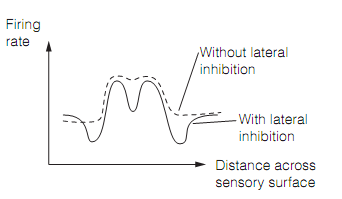
Figure: Contrast enhancement in the presence and absence of the lateral inhibition.