Intracellular recording
Being able to measure membrane potentials straight is crucial to understanding how excitable cells function. A standard method for doing these in individual cells is intra- cellular recording.

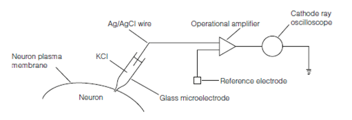
Figure: The circuitry used for intracellular recording.
To record the potential variation across a membrane it is must to have two electrodes, both connected to a voltmeter of some description one inside the cell, the other outside. Because neurons are small the tip of the intracellular electrode impaling the cell required to be very fine. To get this, glass micropipettes are manufactured to have a tip diameter of less than 1 µm. The micropipette is full with an electrolyte (generally KCl at a concentration of 0.15–3 M) to hold the current, so forming the microelectrode. Usually transmembrane potentials are less than 0.1 V and so necessary be amplified with an operational amplifier. From both the intracellular microelectrode has inputs which impales the reference and the cell (bath or indifferent) electrode, that is placed in the solution bathing the cell. If no potential difference exists among the microelectrode and the reference electrode the amplifier output will be zero. If a potential difference exists among the electrodes, moreover, the amplifier generates a signal, the magnitude of that is proportional to the potential. The output of the amplifier goes to the analog-to-digital port of a computer running software that allows display, storage and analysis of the data.