Structure of the blood–brain barrier
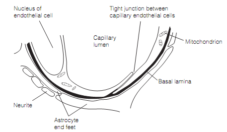
The blood–brain barrier administers what crosses from the blood into the brain extracellular fluid. The anatomical barrier is given by brain capillary endothelial cells that are coupled to each other by tight junctions which are one hundred-fold tighter than is typical for other capillaries. Even small ions will not permeate between endothelial cells in the brain capillaries. Furthermore, the brain capillary endothelial cells display little pinocytosis or receptor-mediated endocytosis. The Brain capillaries are completely covered by the astrocyte endfeet that secrete growth factors (example, angiopoietin 1) which promote the efficacy of tight junctions as shown in figure.

Figure: The structural features of blood–brain barrier. The barrier is formed by the tight Junctions between the endothelial cells.
The circumventricular organs (CVOs), for illustration, the posterior pituitary, lie on the blood side of the blood–brain barrier. They are remote from the rest of the brain by ependymal cells which are coupled altogether by tight junctions. The lack of a blood–brain barrier at the posterior pituitary allows oxytocin and vasopressin to be secreted directly into the systemic circulation. The other CVOs permit the brain to monitor osmolality, or the concentrations of particular ions or molecules for homeostatic functions.