Transmission Electron Microscopy
A beam of electrons is focused by using electromagnetic lenses in electron microscopy. The specimen is mounted within a vacuum so that by atoms the electrons are not absorbed in the air. In transmission electron microscopy, through a thin section of the specimen the beam of electrons is passed that has been stained with heavy metals. The electron-dense metals scatter the incident electrons, thus producing an image of the specimen.
On the contrary with light microscopy where optical lenses focus a beam of light electromagnetic lenses focus a beam of electrons, in electron microscopy. Reason behind it is that electrons are absorbed by atoms in the air; the specimen has to be mounted in a vacuum within an evacuated tube. With biological materials the resolution of the electron microscope is at best 0.10 nm .A beam of electrons is directed in transmission electron microscopy through the specimen and electromagnetic lenses are used to focus the transmitted electrons to generate an image either on a viewing screen or on photographic Film. Thin sections of the specimen are viewed as in standard light microscopy. Yet, for transmission electron microscopy the sections ought to be much thinner (50-100 nm thick). As electrons pass uniformly through biological material, unstained specimens give very poor images. Hence, the specimen ought to be routinely be stained in order to scatter some of the incident electrons by the electromagnetic lenses which are then not focused and so do not form the image. Heavy metals like osmium and gold are frequently used to stain biological materials.Osmium tetroxide preferentially stains certain cellular components, in particular like membranes, which appear black in the image. The transmission electron microscope has adequately high resolution that it may be used to get information regarding the shapes of purified proteins, subcellular organelles. and viruses . In a similar way Antibodies may be tagged with electrondense gold particles to being tagged having a Fluorescent compound in Fluorescence microscopy, and then bound to particular target proteins in the thin sections of the specimen. In the electron microscope when viewed, due to the gold particles small dark spots are seen in the image wherever an antibody molecule has bound to its antigen and so the technique may be used to localize particular antigens.
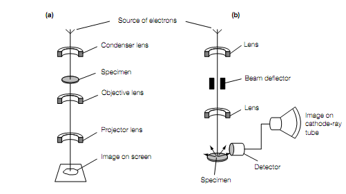
Principal features of (a) a transmission electron microscope and (b) a scanning electron microscope.