Transduction in otolith organs
The macula is an epithelial sheet of supporting cells, and sensory hair cells. Each hair cell is innervated at its base by two nerve fibers, a vestibular afferent and an efferent. The apical border of a hair cell has a single motile kinocilium, that resemble a cilium, and 40–100 stereocilia, microvilli that are progressively shorter the further they are from the kinocilium which is as shown in figure. This defines an axis of polarity for a hair cell, with a direction going away from the minimum stereocilium to the kinocilium. The Stereocilia lying along this axis are linked at their tips by thin filaments called tip links. Stereocilia are the mechanosensory organelles of the inner ear since their tips contain stretch-activated potassium channels that are regulated by the tip links.
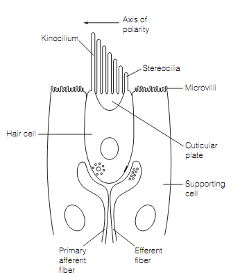
Figure: The hair cell of an otolith organ surrounded by supporting cells of the sensory epithelium.
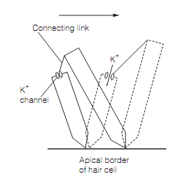
The kinocilium and stereocilia are embedded in a gelatinous matrix that is the otolith membrane, having tiny crystals of calcium carbonate, known as otoliths. The hair cell is atrest if no force acts on the otolith membrane to cause the stereocilia to pivot. In this state the tension in the tip links as shown in figure connecting adjacent stereocilia is slight so only about 10% of the potassium channels gated by them are open, causing a small depolarization.
This is sufficient to sustain tonic release of glutamate, which maintains baseline firing of the main afferent. The Head tilt in the direction of axis of polarity causes the otolith membrane to pull on the stereocilia, making them pivot, therefore increasing the tension in the tip links. This opens stereocilia K+ channels, allowing potassium influx to depolarize the hair cell, increasing glutamate release and raising the afferent firing rate. Tilt in the oppo-site direction reduces tip link tension so K+ channels close, the hair cells hyperpolarize and primary afferent firing drops. Tilt which is perpendicular to the axis of polarity of a hair cell has no effect because stereocilia are not linked in this direction. The tilts in intermediary directions cause graded receptor potentials. The responses of separate otolith afferents are proportional to tilt angle and adapt only with prolonged stimulation. The axes of hair cells are orientated in an orderly pattern so a given stimulus will depolarize some hair cells and hyperpolarize others.