The gram-negative outer membrane
In contrast to the Gram-positive Bacteria the model Bacterium E. coli does not present a coat of peptidoglycan to the outside world in Figure 1. Alternative the murein is suspended in the periplasmic space and there is a second outer membrane of LPS. The Gram-negative outer membrane has some features in common with the cytoplasmic membrane in which it is a lipid bilayer and is considered to be a fluid mosaic. However, rather than being composed of phospholipid alone there are several lipids with polysaccharide attached. This alters the physical and chemical properties of the membrane in which it is much more porous to much larger molecules. This porosity is enhanced through the presence of several transport and porin proteins.
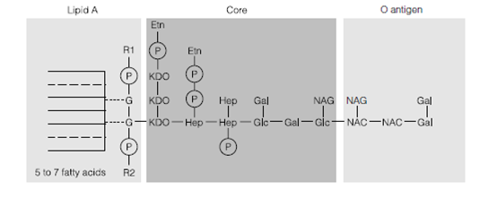
Our detailed knowledge of the outer membrane structure of Gram-negative Bacteria has been driven through the antigenic properties of LPS so the details purporting to represent Gram-negatives are in reality more relevant to pathogenic enteric Bacteria like as E. coli, Salmonella and many more. The presence of LPS can provoke a strong immune response in mammals so in genera including Salmonella, Escherichia, Pseudomonas, Shigella, Neisseria and Haemophilus the term LPS has become interchangeable with endotoxin. Chemistry of this LPS outer membrane is variable and complex among species. In Salmonella the lipid part of the molecule is phosphorylated glucosamine linked to long carbon chains that in contrast to membrane lipids may be branched. Lipid a of Gram-negative Bacteria is covalently linked to the core polysaccharide in Figure 3 but even if separated from the rest of the molecule it is frequently toxic to mammals even if the bacterium of origin is nonpathogenic. The Lipid A serves to anchor the LPS to the rest of the outer membrane.
The core polysaccharide or R antigen or R polysaccharide is the similar in all members of a particular genus and is made up of hexose and heptose sugars plus ketodeoxyoctonate. Lastly the O antigen or somatic antigen or O polysaccharide is made up of three to five repeating sugars. The composition of the repeat varies from species to species and can be repeated from 1 to 40 times. This is the major antigenic determinant of pathogenic cells.

Figure . Lipopolysaccharide. P, phosphate; G, glucosamine; NAG, N-acetylglucosamine; NAC, N-acetylgalactosamine; Gal, galactose; Glc, glucose; Hep, heptose; Etn, ethanolamine; R1 and R2, phosphoethanolamine or aminoarabinose; KDO, ketodeoxyoctonate.