Gross anatomy
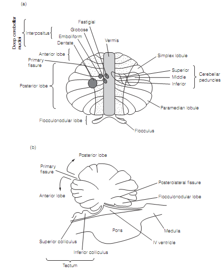
The human cerebellum is around one quarter of the mass of the brain and is divided into three lobes, the anterior lobe and posterior lobe, divided by the primary fissure, and the flocculonodular lobe, estranged from the posterior lobe by the posterolateral fissure as shown in figure. The lobes are further subcategorized into lobules that are differently named in humans and other mammals. Longitudinally cerebellum has a central vermis and two lateral hemispheres.
Over the surface of the cerebellum lies the cerebellar cortex that is folded into coronal strips known as folia. The cerebellum has an internal core of white matter having deep (intracerebellar) nuclei. These, altogether with the vestibular nuclei, give the output of the cerebellum. Efferent and afferent connections of the cerebellum go by way of three pairs of the cerebellar peduncles.

Figure: Cerebellar anatomy. (a) A diagram of the cerebellum unfurled and viewed from above. Positions of the deep cerebellar nuclei are shown on the left and of the cerebellar peduncles on the right. The medial zone is pale gray, the intermediary zone stippled, and lateral zone is clear. (b) Sagittal section via the brainstem and cerebellum.