Striatum
The putamen and caudate nucleus are functionally a single unit, the dorsal striatum though are divide anatomically by the internal capsule. The adjoining ventral striatum (i.e., nucleus accumbens), that is part of the limbic system, has same circuitry.The striatum obtains excitatory input from the cortex through the glutamatergic corticostriate pathway as shown in figure. The input is prearranged topographically therefore somatotopy is conserved in the projections from the motor cortex and somatosensory cortex. The Corticostriate axons terminate on the main neuron kindin the striatum, the medium spiny neuron. This build up 95% of striatal neurons, uses GABA as their transmitter, and give the inhibitory outcome of the striatum. There are two populations of the medium spiny neurons with various connections and neurochemistry. One kind has substance P (SP) and dynorphin (DYN) as co-transmitters, state dopamine D1 receptors, and plans to the globus pallidus pars interna (GPi) & substantia nigra pars reticulata (SNpr). The second uses enkephalin (ENK) as a co-transmitter, expresses D2 dopamine receptors, & plans to the globus pallidus pars externa (GPe).
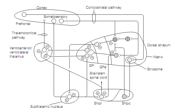
Figure: Connections of the basal ganglia.
The medium spiny neurons obtain projections through the nigrostriatal pathway from the substantia nigra pars compacta (SNpc). The SNpc uses dopamine as transmitter. Since the two kinds of medium spiny neuron express various dopamine receptors they are in a different way modulated by this input. At the GABA/SP/DYN cells, dopamine performing on D1 receptors improves the consequence of excitatory cortical input. In contrary, the action of dopamine on D2 receptors on GABA/ENK cells is to decrease the consequence of cortical excitation. Medium spiny neurons contain third input from large aspiny interneurons which constitute around 2% of striatal neurons. Such cells use ACh as a transmitter, are excitatory, and driven by the cortical inputs.
Staining of the striatum for acetylcholinesterase illustrates it to be compartmented into a heavily stained matrix and a lightly stained 3-dimensional labyrinth, the striosomes, which are around 10–20% of the striatal bulk. The connectivity of cells in these compartments is diverse. The matrix gets inputs from throughout the cerebral cortex and sends outcomes to the whole globus pallidus and SNpr, while the striosomes get limited input from the prefrontal cortex and plan to the SNpc. The matrix is concerned with sensorimotor function, whereas striosomes are related with the limbic system, and might control the dopaminergic pathway from SNpc to striatum.