Overview Anatomy of the basal ganglia
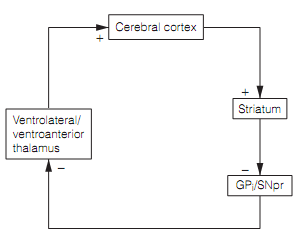
The traditional role of the basal ganglia is in selecting suitable learned motor series, and in stimulus–reward learning to form the novel motor strategies. They consist of numerous extensively interconnected structures, the striatum (i.e., the caudate and putamen), the globus pallidus (i.e., pars interna & pars externa), the substantia nigra (i.e., pars compacta & pars reticulata), and the subthalamic nucleus. Nearly all inputs to the basal ganglia are from the cerebral cortex and go through the striatum. The outcome of the basal ganglia appears from the pars interna (i.e., internal section) of the globus pallidus, and substantia nigra pars reticulata, to go to the thalamus. The thalamus plans back to the cortex therefore closing a loop. The thalamocortical axons turn to the similar area of cortex that gave rise to the striatal inputs as shown in figure below.

Figure: Block diagram of the interface among basal ganglia and cerebral cortex. Here, GPi=globus pallidus pars interna; SNpr=substantia nigra pars reticulata.
The motor basal ganglia circuitry is dependable for the execution of suitable preprogrammed motor series during voluntary movements. Typically the basal ganglia constitute portion of the extra-pyramidal system, on the basis which lesions of the basal ganglia generate quite various symptoms from lesions of the corticospinal tract.