Hypothalamic–pituitary connections
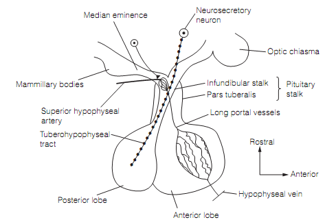
The pituitary gland is splitted into the neurohypophysis and the adenohypophysis. The neurohypophysis, that is a direct outgrowth of the hypothalamus, consists of the posterior lobe, the infundibulum, and the median eminence. The adenohypophysis have anterior lobe, an intermediary lobe (badly developed in humans), and the pars tuberalis (an expansion surrounding the infundibulum). The pars tuberalis and the infundibulum altogether make up the pituitary stalk as shown in figure below.

Figure: The pituitary showing its neural and vascular connections with the hypothalamus. In humans the tuberohypophyseal tract consists of around 100 000 axons.
Most of the endocrine and autonomic functions of the hypothalamus include the paraventricular nuclei (PVN). These contain two classes of peptide-secreting neuroendocrine cells:
1. Magnocellular (i.e., large) cells that send their axons via the median eminence down the infundibular stalk into the posterior lobe as the tuberohypophyseal tract. Hormones are made in the cell bodies of the magnocellular cells and elated down their axons for discharge in the posterior lobe.
2. Parvocellular (i.e., small) cells have brief axons that terminate on capillaries in the median eminence. These capillaries drain into long portal vessels which descend to form venous sinusoids in the anterior lobe; this vascular bed is the portal system. Hormones secreted by the parvocellular neurons into the median prominence are carried through the hypothalamic-pituitary portal circulation to the anterior lobe.
Therefore the hypothalamus has a neural link to the posterior lobe, though a vascular link to the anterior lobe. Both lobes have fenestrated capillaries which lie on the blood side of the blood–brain barrier which drains into the systemic circulation. By this route the pituitary hormones are delivered to the body.