Cholinergic pathways
The motor neurons in motor nuclei of cranial nerves and ventral horn of the spinal cord are cholinergic, as preganglionic autonomic neurons, and the axons of all such cells project into the peripheral nervous system. The Cholinergic interneurons are present in the striatum and the nucleus accumbens.
Three areas within the brain hold cholinergic neurons which project centrally. Mainly caudal are those of the pontine reticular formation which send axons into the spinal cord or forward to the thalamus, amygdala, and basal forebrain. These are significant in regulating sleep and wakefulness.
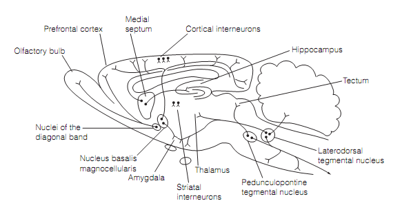
The second area, the basal forebrain, holds the nucleus basalis of Meynert (nBM) that projects extensively to the cerebral cortex. The third area, the medial septum, gives increase to the septohippocampal pathway as shown in figure. In primates, the cholinergic neurons of the nBM show short changes in firing rate during the behavioral tasks, mainly when reinforcing stimuli are presented. The Lesions of nBM generate impairment in recall for tasks learnt before the surgery and in acquisition of the new learning. The ACh generates long term facilitation of the neurons in neocortex and hippocampus by acting at muscarinic receptors to close potassium (KM) channels, making the cells more probable to fire in response to the excitatory inputs. Therefore the forebrain cholinergic system appears to be a selective arousal system, activate by satisfying or salient events that facilitates the learning.

Figure: Major cholinergic pathways in a sagittal part of the rat brain. Nucleus basalis magnocellularis of the rat is termed as the nucleus basalis of Meynert in primates.