Q. What is Pulmonary Edema?
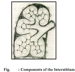
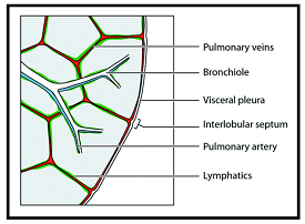
When the capillary pressure exceeds the plasma osmotic pressure, fluid first accumulates in the interstitial spaces. The components of the interstitium (central and peripheral) are shown in Fig.
The central interstitium invests the bronchovascular bundle and extends from centre to periphery. Fluid accumulating in this perivascular and peribronchial interstitium causes an apparent increase in the size of vessels at the hilum, as well as loss of definition of vessels on the CXR.
The peripheral interstitium consists of subpleural, inter and intralobular septal components. One of the early manifestations of interstitial edema on the CXR are septal lines, commonly Kerley B lines. These are short, straight, horizontal lines, best seen in the lower zones, representing thickening of the interlobular septae (Figure).

Other lines described are Kerley A lines (4 to 6 cm, radiating from the hilum, more in the upper zones) and Kerley C lines (short, crisscrossing lines), all representing thickened interstitium. Further fluid accumulation results in edema of alveolar walls and alveolar edema. This characteristically has an "air-space" appearance with coalescent pulmonary opacities, resembling cotton wool. Air space opacification creates a contrast between air filled bronchi and the surrounding lung, and this may produce an air-bronchogram. Typically, there is a perihilar distribution, resulting in a "bats wing appearance". Rapid clearing with antifailure measures is seen.
