Reference no: EM132372213
Virtual Microscope Lab - Cheek Cells
You will be using a "virtual microscope" which can be accessed on the internet. In a classroom lab, we would obtain cheek cells by scraping the inside of the mouth with a toothpick and then rubbing the toothpick on a drop of water with blue stain. The blue helps you see the cells which are normally a clear color. The virtual lab begins at the step where you place the slide on the microscope stage.
Access the Virtual Microscope and watch the introductory video, and then click on the link that says "the virtual scope."
1. Familiarize yourself with the microscope, run the tutorial and examine the parts you will be working with. The lens in the eyepiece magnifies 10 times, so multiply that by the magnification of the objective used to get total magnification.
2. View the slide labeled cheek smear. Sketch the image at Scanning, Low and High Power. LABEL on high power the CELL MEMBRANE, CYTOPLASM, and NUCLEUS.
3. Use the internet or your textbook to define or describe each of the following terms that relate to the cell.
eukaryote
nucleus
cell membrane
cytoplasm
4. The mouth is the first site of chemical digestion in a human. Your saliva starts the process of breaking down the food you eat. Keeping this in mind, what organelle do you think would be the most numerous inside the cells of your mouth? (Hint: what organelle is responsible for breaking things down and digesting?)
5. What is the function of chloroplasts?
6. Name two structures found in plant cells but not animal cells.
7. Name three structures found in plant cells AND in animal cells.
8. What structure surrounds the cell membrane (in plants) and gives the cell support.
Procedure: Go to plantcells (see attached file) which contains images of cells as they were viewed in a lab. You will use these images to complete this worksheet.
Part A - Onion Cells
View under the microscope and sketch the cells at each magnification. Label the cells as they appear under high power.
Part B - Elodea Cells
View and draw the prepared slides of elodea (anacharis), which is an aquarium plant. As the slide warms from the light of the microscope, you may see the chloroplasts moving, a process called cytoplasmic streaming.
9. Describe the shape and the location of chloroplasts.
10. Why were no chloroplasts found in the onion cells? (hint: think about where you find onions)
11. Which type of cell was smaller - the onion cells or the elodea cells?
12. Fill out the Venn Diagram below to show the differences and similarities between plant cells and animal cells. The similarities will be in the center overlap and the differences will be on each side.
Cell Structures - Animal Cell - Label numbered parts
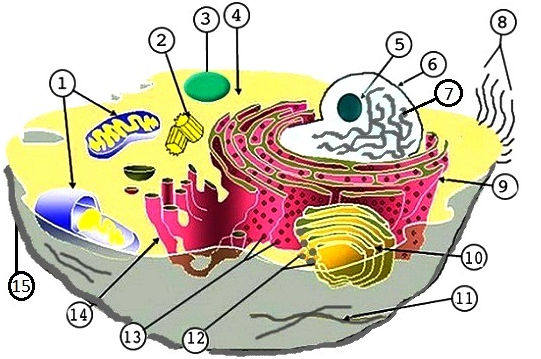
1. ___________________________
2. ___________________________
3. ___________________________
4. ___________________________
5. ___________________________
6. ___________________________
7. ___________________________
8. ___________________________
9. ___________________________
10. ___________________________
11. ___________________________
12. ___________________________
13. ___________________________
14. ___________________________
15. ___________________________
16. Which organelle contains its own DNA?
17. What is the difference between smooth and rough ER?
Cell Structures - Plant Cell - Label lettered parts

A. ___________________________
B. ___________________________
C. ___________________________
D. ___________________________
E. ___________________________
F. ___________________________
G. ___________________________
H. ___________________________
I. ___________________________
J. ___________________________
K. ___________________________
L. ___________________________
M. ___________________________
N. ___________________________
Attachment:- Virtual Microscope Lab Assignment File.rar