Reference no: EM13494626
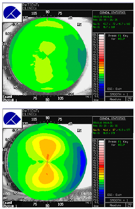
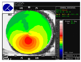
Topography maps are shown. For comparison a map from two normal patients are shown. The patient is diagnosed as having Keratoconus.


Questions
1. How does topography work?
2. Why does the normal cornea flatten towards the periphery?
3. Where is the apex of the cone?
4. What are the treatment options for keratoconus? Explain how each could improve vision, their limitations, and possible complications.
A few years later, the left eye becomes more astigmatic and shows signs of keratoconus.. The right eye progressed and eventually required a corneal transplant. This worked for about 15 years when signs of ectasia began to appear again.
5. What is the significance of the disease being bilateral in terms of understanding its cause?
6. How could the donor cornea, which was normal, begin to show signs of keratoconus?
7. Describe the current hypotheses for the cause of Keratoconus. (You need to do careful Pubmed searches).