Reference no: EM13793147
Stripping Analysis of Metals in Industrial Samples
Objective:
(1) The principle behind stripping analysis and the concept of preconcentration.
(2) Use of a dropping mercury electrode and differential pulse voltammetry.
(3) Use of the standard addition method for quantification.
Principles
Stripping analysis (SA) is a method for elemental analysis that utilizes a bulk electrolysis step (deposition) to preconcentrate a substance from solution into or onto an electrode. After the deposition step, the material is redissolved (stripped) from the electrode using some voltammetric methods. Different analytes (metals) are oxidized (stripped) at different potentials which can be employed for the qualitative identification of analyte. If the conditions during the deposition step are maintained constant, exhaustive electrolysis of the solution is not necessary and, by proper calibration and with fixed electrolysis (deposition) times, the stripping peak currents can be employed to find the analyte concentration. The following shows the reactions related to SA of Cd2+ using a Hg electrode.
(1) Cd2+ (solution) + 2e- + Hg = Cd(Hg) preconcentration at the Hg electrode
(2) Cd(Hg) (at the electrode) - 2e- =Cd2+ anodic stripping (reoxidation)
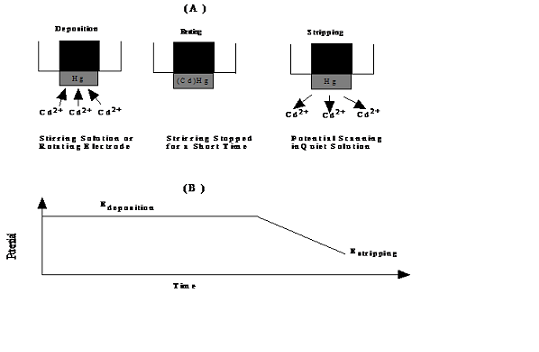
In step (1), metal ions to be analyzed in solution are reduced at a mercury electrode in the hanging mercury drop mode (SMDE) by applying an enough negative potential. In step (2), a voltammetric technique, viz., differential pulse voltammetry (DPV), will be used to achieve the metal stripping (oxidation) . In DPV, the potential at the working (analyzing) electrode is varied linearly with time. At the same time, a square wave with a small pulse (typically 50 mV). By scanning the potential from low to high volts, the preconcentrated metals at the electrode will be stripped off sequentially depending on its oxidation power. The overall processes are shown in the schematic Figure 1 below.
Instrumentation: A CHI615 Electrochemical Workstation will be used to obtain the stripping voltammograms. A glass electrochemical cell ("shot glass") housing a dropping mercury capillary, a Pt counter or auxiliary electrode, and a Ag/AgCl reference electrode will be used. A stirring bar will be employed for achieving higher preconcentration efficiency.
Solutions and Materials:
(A) A 10-mL unknown solution (unknown metal dissolved in 1% HNO3 solution) will be provided to each group.
(B) Three standards of Cu2+, Pb2+, and Cd2+ (1000 ppm each) will also be provided.
CAUTION: Hg is highly toxic and very difficult to clean if spilled. Be extremely careful in not breaking the capillary and properly dispose the Hg waste at the bottom of the glass cell (shot glass). To maintain an intact Hg capillary, when you need to dispose the sample solution and Hg droplets in the cell, please use one hand to hold the glass cell while use your other hand to swing the stirrer (glass support) to your right. Carefully lower the glass cell vertically until you can see the tip of the capillary not be crashed with the glass cell wall.
To dispose the Hg droplets into the large plastic container next to the instrument without losing the small stirrer bar, please use the large magnetic stirrer bar to take the small bar out of the solution first. You can then dump the solution together with the Hg droplets into the waste container.

Figure 1. Schematic Representation of the Steps Involved in a SA Experiment (Top Panel) and the Applied Potential during the Experiment (in the example the pulses in DPV superimposed on the linear scan are not shown)
Suggested Procedure:
(1) Clean the glass cell, the capillary and auxiliary electrode by squirting deionized water from your wash bottle. Carefully remove the glass cell and wipe its interior with tissues. Add the entire 10 mL unknown solution into the glass cell and put it back to the apparatus. Now insert the Ag/AgCl electrode through the hole on the right of the cap.
(2) Be sure that the toggle switch on the top is at "SMDE" and the "Size" is selected as "8". Leave the stirring rate in the middle panel at the preset value.
(3) Connect the green lead from the Electrochemical Analyzer to the horizontal pin on the side of the Hg pool, the red lead to the Pt auxiliary electrode, and the white lead to the Ag/AgCl reference electrode.
(4) O2 dissolved in the sample solution interferes with the measurement and needs to be removed. This is accomplished by degassing the solution with N2. On the front panel of the Controlled Growth Mercury Electrode device, slowly turn the knob counterclockwise (from "Close" to "Open") until you can see a stream of gas bubbles purging through the sample solution. Please do not open the knob too widely as the high pressure of the purging N2 gas will splash your sample solution. Degas for 5 min.
(5) Click on the icon "Chi610a.exe".
(6) Click on the button of "Technique" (). Then, select "Differential Pulse Voltammetry" and click on "OK".
(7) Click the icon next to the T (Parameters/) to set the parameters.
(8) Put the value of initial E (V) of -0.8, Final E (V) of 0.2, Quiet Time (Sec) of 2, and Sensitivity (A/V) of 1.e-006. Click on "OK".
(9) Go to "control," click on "stripping mode" and enable "Stripping Mode Enabled,""Purge During Deposition,"and "Stir During Deposition", enter 10s in the "Deposition Time". Then, click "OK".
(10)Click the "Run" bottom ().
(11)The instrument will count the deposition time. Then collecting the voltammogram.
(12)Go to "Graphics", choose "Graph Options".
(13)Find "Header": type in a descriptive title, such as "Unknown Run 1," then, click on "OK".
(14)Go to "File", click on "Save As", type in a descriptive file name, and select to store in a proper folder, then click on "Save".
(15)Click "Run" button to run again.
(16)Repeat the same procedure: Add the graph a header, and save with different file name in a correct folder.
(17)Go to "Graphics", select "Overlay Plots".
(18)Click to select the first run as the first corresponding file, click on "Open". If the two curves are highly comparable, you can do the third run.
(19)After the third run, to gauge the reproducibility, overlay the first and second runs by the same procedure.
(20)If the 3 curves are reproducible (overlayable), go to the next step. If not, run the fourth time.
Time Dependent Study
(21)Go to "control," click on "stripping mode" and enable "Stripping Mode Enabled,""Purge During Deposition,"and "Stir During Deposition", enter different times in the "Deposition Time" (5, 10, 20, and 30 (s))for different runs. Then, click "OK".
(22)Click on Triangle shaped "Run" button.
(23)Use the "Overlay Plots" function to obtain a reproducible stripping voltammogram contains 2 runs for each deposition time.
Data Analysis
(24)To determine peak high/peak area, open the file you want to analyze, click on icon "Data Plot" ().
(25)The program may automatically identify the "Peak Potential (Ep)", "Peak Current (ip)", and "Peak Area (Ap)". The values are shown in blue at the bottom right corner.
(26)If the peak(s) is/are not identified, click on "Manual Results" (), an upward arrow appears.
(27)Along the baseline of unidentified peak, use a tip of the arrow to draw a line across the unidentified peak until the line meets the baseline on the other side of the peak.
(28)The values corresponding to the unidentified peak(s) show on the right hand side in blue.
(29)Record the values on you lab notebook.
(30)Study the deposition time dependence of the stripping peak currents (e.g., is the peak current proportional to the deposition time?).
(31)Next you are ready to quantify the concentrations of Cu2+, Cd2+ and Pb2+ in the unknown sample. This is to be accomplished by the method of standard addition (addition of a small amount of highly concentrated standard to the unknown sample to record an increase in the signal). First, make sure that the deposition time in the program is selected as 10 s. Pipet 10 µL of 1000 ppm Cd2+ with a metal-free pipet into the solution through the hole on the cap of the glass cell. Be certain that you have dislodged the small drop at the end of the pipet tip into the sample. This is followed by adding 10 µL each of the Cu2+ and Pb2+ standards. Degas the solution again with N2 for 3 min.
(32)Collect three reproducible stripping voltammograms.
(33)Next, add additional 10 µL of the standards into the unknown and collect again three reproducible stripping voltammograms.
(34) If time allows, you can add another 20 µL of the standards and collect three voltammograms.
(35) By either using the equation of the method of standard addition or plots in Excel, you will be able to quantify the unknown Cu2+, Cd2+ and Pb2+concentrations.
(36) Dispose the sample solution together with the Hg waste into the waste bottle (cf. procedure under Caution).
(37) You can convert the data file into a text format for you to plot representative voltammogram(s) in Excel. Under "File", select "Convert to Text" and the program will prompt you to open a file with extension of "bin". After conversion, the file will have the same name but with an extension of "txt", which is importable into Excel.
Questions:
(1) What is the relationship between the stripping peak current and the scan rate in LSV and that between the peak current and the deposition time?
(2) What is the reproducibility of your stripping analysis experiment?
(3) Report the concentration (in ppb or ppm) of Pb2+ in the unknown sample.
(4) Discuss the factors determining the reproducibility of SA.
(5) Speculate the advantages and disadvantages of in-situ and ex-situ Hg films.
(6) What affects the detection limit of the SA method?