Inner ear anatomy
The auditory section of the inner ear is the cochlea, a bony canal 3.5 cm long, that spirals two and three quarter turns around a central pillar, the modiolus. In the cochlea lies a tubular extension of the membranous labyrinth, the cochlear duct, joined to the modiolus and the outer wall of the cochlea. This splits the cochlea into three compartments, the scala media that contains endolymph, and the scala vestibuli and the scala tympani that contain perilymph and are continuous with each other through a small gap termed as helicotrema located at the apex of the cochlea where the cochlear duct ends blindly as shown in figure below:
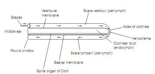
Figure: The cochlea, depicted unfurled. Arrows represent the direction of propagation of sound waves via the perilymph.
Pressure waves produced at the oval window are propagated via the scala vestibuli into the scala tympani and to the round window where the energy disperses. During their passage the pressure waves cause oscillations of the basilar membrane, the floor of the scala media on that rest the sensory apparatus, the spiral organ of Corti as shown in figure below:
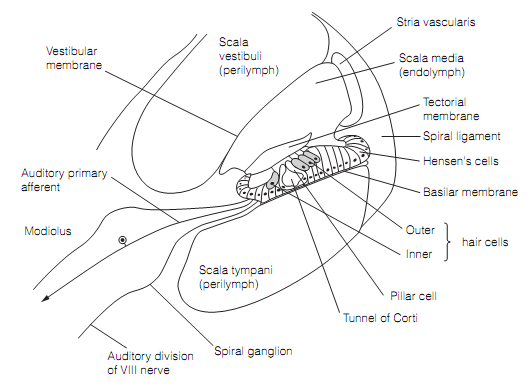
Figure: Transverse section through the cochlea displaying the organ of Corti.
The spiral organ is a narrow sheet of columnar epithelium running the length of the cochlear duct. The epithelium holds sensory hair cells resembling those in the vestibular apparatus. Single row of 3500 inner hair cells form ribbon synapses with myelinated axons of large bipolar cells (type I) in the spiral ganglion of the cochlear nerve. Each inner hair cell is innervated by around10 such axons, a large degree of divergence. There are about 12 000 outer hair cells arranged in 3 rows. These are innervated by an unmyelinated axon from small bipolar cells (type II) in the spiral ganglion, each one synapses with 10 hair cells, representing significant convergence.